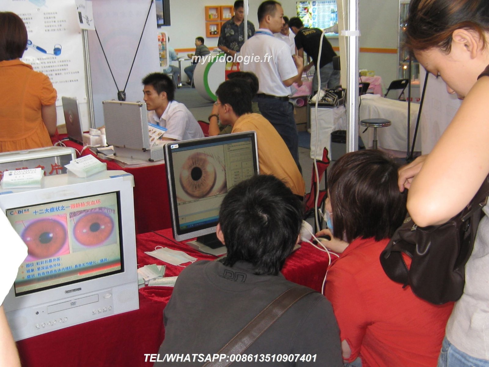
Professional practitioners examine these iris zones to identify potential respiratory concerns before symptoms manifest. This detailed examination process combines traditional iridology principles with modern diagnostic approaches.
Understanding left eye iridology chart interpretation requires knowledge of specific sector locations and their corresponding organ relationships. Each zone tells part of your respiratory health story.
Iris Sector Fundamentals
The human iris divides into twelve major sectors, similar to clock positions. Each sector corresponds to specific organs and systems throughout your body.
Practitioners use the iridology chart left eye as their primary reference tool. This chart maps precise anatomical relationships between iris zones and body areas.
The left eye iridology chart reveals your right side body structures. This mirror relationship forms the foundation of iris interpretation principles.
The Mirror Principle in Iridology
Your left eye reflects your right side organs. Your right eye shows left body structures.
This crossover pattern applies consistently across all iris mapping systems. Practitioners worldwide recognize this fundamental relationship.
The right lung appears in the upper-left quadrant of your left iris. This specific sector occupies approximately the 2-3 o’clock position.
Historical Development of Iris Mapping
Early iridology pioneers established sector divisions through clinical observation. Dr. Ignatz von Peczely documented initial iris-organ correlations in 1881.
Bernard Jensen refined these charts throughout the 20th century. His work created standardized zones used by modern practitioners.
Contemporary iris analysis builds upon decades of documented case studies. These historical findings provide the framework for current interpretation methods.
Professional Insight: Accurate iris sector identification requires proper lighting and magnification equipment. Many experienced practitioners use specialized iridology cameras to capture detailed iris images for thorough examination. Advanced software tools can help identify subtle patterns and changes that might be missed during visual inspection alone.

Anatomical Location of Right Lung Sectors
The right lung sector occupies a specific triangular area within your left iris. This zone extends from the pupil margin outward toward the iris periphery.
Precise location falls between 2 o’clock and 3 o’clock positions. The sector width spans approximately one-twelfth of the total iris circumference.
Practitioners examine three distinct layers within this respiratory zone. Each layer provides different information about lung tissue health.
Zone Boundaries and Landmarks
The inner boundary begins at the autonomic nerve wreath surrounding your pupil. This ring separates the pupil zone from the body zone.
The outer boundary reaches the iris periphery where pigment density typically increases. This outer ring represents the skin and circulation zone.
Vertical landmarks help pinpoint exact respiratory areas. The 2:30 position typically corresponds to upper right lung lobes.
Three-Dimensional Sector Structure
The humoral zone (inner third) reflects body fluid balance affecting lung tissue. Darkness or cloudiness here may indicate accumulated toxins.
The structural zone (middle third) shows actual lung tissue integrity. Lacunae or fiber patterns in this area warrant closer attention.
The circulation zone (outer third) reveals blood and lymph flow to respiratory organs. Color variations and ring formations appear most prominently here.
Primary Right Lung Sector Features
- Location: 2-3 o’clock position in left iris
- Width: Approximately 30 degrees of arc
- Depth: Three distinct tissue layers
- Adjacent sectors: Thyroid (above), bronchi (below)
- Key structures: Upper, middle, and lower lobe representations

Correlation with Respiratory Anatomy
The upper portion of the lung sector corresponds to the superior lobe. This area extends highest in the sector triangle.
The middle area represents the middle lobe, unique to the right lung. This zone appears in the central sector region.
The lower portion indicates the inferior lobe and diaphragmatic surface. This section lies closest to the 3 o’clock boundary.
Iris Patterns as Lung Health Indicators
Specific iris patterns within the lung sector provide valuable health insights. Practitioners analyze fiber density, color variations, and structural formations.
Normal healthy tissue displays consistent fiber patterns with uniform coloration. Disruptions to these patterns may indicate developing concerns.
The following patterns appear most frequently during lung sector examinations. Each pattern carries specific interpretive significance.
Lacunae and Crypts in Respiratory Zones
Lacunae appear as dark openings between iris fibers. These gaps suggest inherent tissue weakness in corresponding organs.
Closed lacunae show honeycomb-like formations with intact fiber borders. Open lacunae reveal deeper pigment loss with ragged edges.
Multiple lacunae within the lung sector may indicate hereditary respiratory weakness. Practitioners note their size, shape, and exact position.
Fiber Density and Texture Analysis
Tight, compact fibers indicate strong constitutional lung tissue. Loose, separated fibers suggest reduced respiratory resilience.
Radial furrows cutting through the lung sector show accumulated body stress. These lines deepen over time with chronic tension.
Wavy or disrupted fiber patterns reflect past inflammation or injury. The area affected corresponds to specific lung regions.
| Pattern Type | Visual Appearance | Potential Interpretation | Clinical Significance |
| Lacunae | Dark holes or gaps in fiber structure | Inherent tissue weakness | Increased susceptibility to respiratory issues |
| Pigment spots | Orange, brown, or yellow deposits | Toxic accumulation | May reflect past infections or environmental exposure |
| White signs | Bright white fiber elevations | Active inflammatory response | Acute or recent lung irritation |
| Radial furrows | Lines radiating from pupil outward | Nerve stress and tension | Chronic stress affecting respiratory function |
| Contraction furrows | Concentric circular grooves | Muscle tension patterns | Breathing restriction or anxiety-related patterns |
Color Variations and Their Meanings
White or silver deposits indicate acute inflammatory processes. These bright areas suggest current tissue irritation.
Yellow or orange tones show accumulated waste materials. This discoloration reflects impaired elimination through lung tissue.
Dark brown or black spots reveal old, encapsulated toxins. These areas represent past exposures or chronic burden.


Interpretation Methodology: Step-by-Step
Systematic examination follows established protocols developed by experienced practitioners. This methodical approach ensures consistent, reliable findings.
Professional iris analysis requires proper documentation and comparison over time. Sequential images reveal progressive changes indicating health trends.
The following methodology represents current best practices in iridology assessment. Each step builds upon previous observations.
Initial Visual Survey
Begin with overall iris observation under consistent lighting conditions. Note general fiber density and dominant coloration.
Identify the autonomic nerve wreath position and integrity. This ring provides crucial orientation for sector location.
Locate the right lung sector precisely at the 2-3 o’clock position. Mentally divide this area into thirds for detailed examination.
Detailed Sector Examination
Examine the inner humoral zone for darkness or cloudiness. Note any pigment deposits or color variations.
Study the middle structural zone for lacunae, fiber disruptions, or unusual patterns. Document size, shape, and location of any formations.
Assess the outer circulation zone for ring formations and color variations. These areas reveal vascular health affecting respiratory tissue.
Professional Tip: Capturing high-quality iris images significantly improves diagnostic accuracy and documentation. Many practitioners maintain detailed photographic records to track changes over months or years. For technical questions about optimal imaging equipment and techniques, professional support is available through specialized iridology equipment providers.
Technical assistance available: WhatsApp: +86 135 1090 7401
Comparative Analysis Process
Compare findings with standard iridology chart left eye references. Cross-reference observed patterns with documented interpretations.
Note bilateral symmetry by examining the corresponding left lung sector in the right eye. Asymmetrical findings warrant additional attention.
Document all observations systematically in client records. Include sketches or annotated photographs for future reference.
- Establish proper lighting and magnification setup
- Capture baseline iris photographs from multiple angles
- Locate and mark the right lung sector boundaries
- Examine each zone layer systematically
- Document all observed patterns, colors, and formations
- Compare findings with reference charts and previous images
- Cross-check observations with client health history
- Generate written report with specific findings
- Schedule follow-up examination to track changes
- Integrate findings with other assessment tools
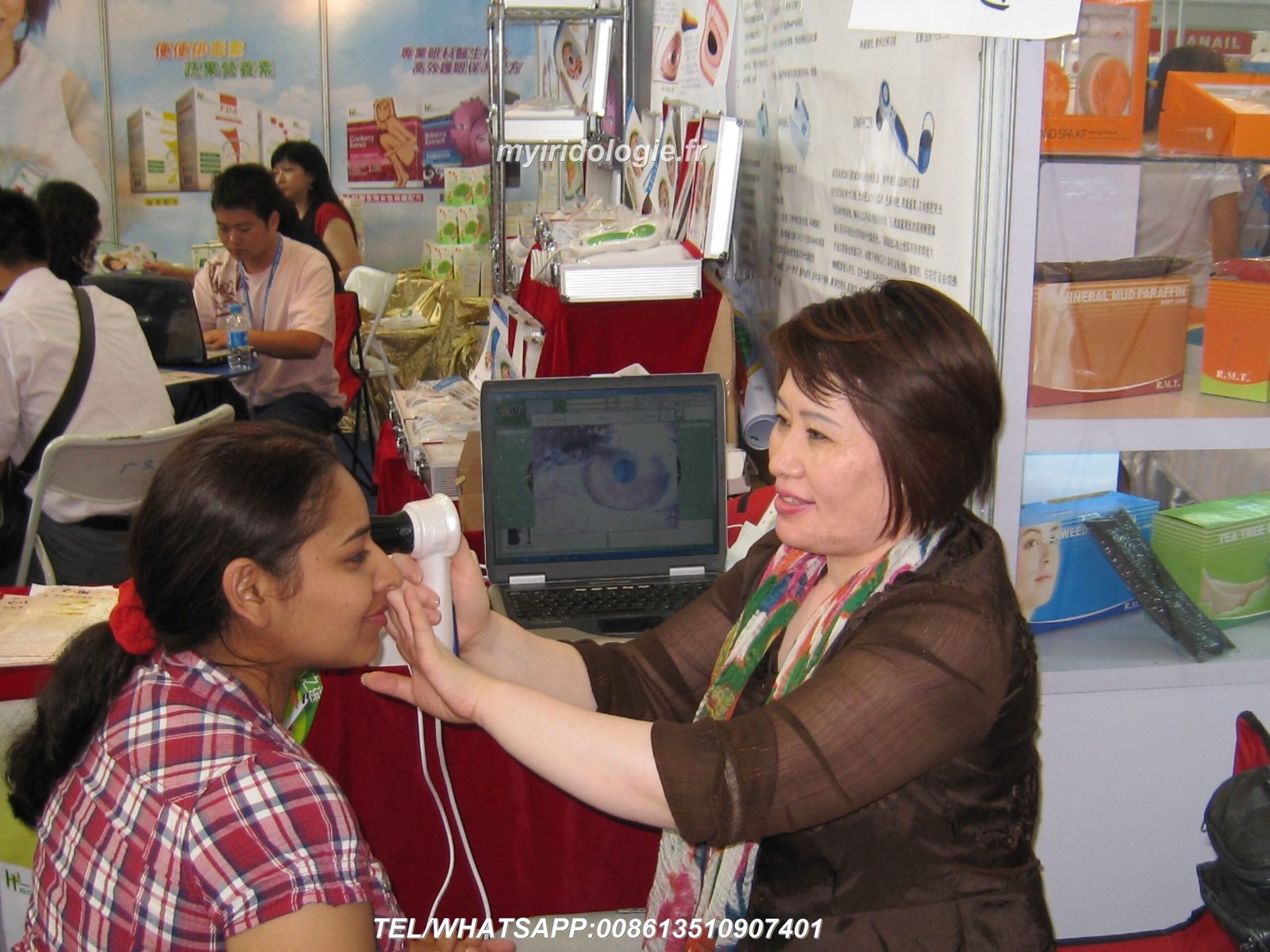

Common Findings and Clinical Significance
Experienced practitioners recognize recurring patterns that appear across many clients. These common findings correlate with specific respiratory concerns.
Understanding typical iris variations helps differentiate normal constitution from developing issues. Context always matters when interpreting iris signs.
The following observations appear most frequently during professional lung sector examinations. Each finding carries interpretive weight within the broader health context.
Stress Rings and Their Impact
Concentric rings encircling the iris indicate chronic tension affecting multiple body systems. These contraction furrows appear as circular grooves.
When stress rings pass through the lung sector, they suggest breathing restrictions. Shallow breathing patterns often indicate accompanying anxiety.
Deep, numerous stress rings reflect long-term body tension. This pattern correlates with reduced respiratory capacity over time.
Lymphatic Rosary Formation
Small white or cream-colored clouds in the outer iris zone indicate lymphatic congestion. This pattern suggests impaired immune response.
When appearing in the lung sector, lymphatic rosary may reflect chronic respiratory mucus. These clouds represent accumulated waste materials.
Clearing these formations requires supporting overall lymphatic drainage. Respiratory health improves as lymphatic circulation enhances.
Acute Respiratory Indicators
- White fiber elevation (active inflammation)
- Bright yellow discoloration (recent infection)
- Vessel tortuosity (circulatory stress)
- Pupil distortion toward lung sector (nerve irritation)
- Increased pigment density (immune response)
Chronic Respiratory Patterns
- Deep lacunae (inherent weakness)
- Orange-brown deposits (old toxic burden)
- Fiber thinning (tissue degeneration)
- Multiple radial furrows (chronic stress)
- Peripheral darkening (circulation deficiency)
Toxic Accumulation Signs
Orange or rust-colored deposits within the lung sector suggest environmental exposure history. These areas may reflect smoking, pollution, or occupational irritants.
Yellow psoric spots indicate sulfur-related toxic burden. This pattern appears commonly in individuals with chronic sinus conditions.
Dark brown or black markings show deeply encapsulated toxins. These deposits represent the body‘s attempt to isolate harmful materials.
Constitutional Weakness Patterns
Large, open lacunae spanning multiple layers indicate significant inherent weakness. These formations suggest reduced constitutional resilience.
Honeycomb patterns with numerous small crypts show moderate genetic predisposition. Supporting lung health becomes particularly important with this pattern.
Fiber separation with visible pigment layers beneath indicates breakdown of tissue integrity. This pattern warrants comprehensive respiratory support strategies.


Professional Tools for Accurate Assessment
Accurate iris examination requires specialized equipment beyond basic visual inspection. Professional-grade tools enable detailed documentation and analysis.
Modern technology has significantly enhanced iridology practice capabilities. Digital imaging and analysis software provide unprecedented detail and tracking abilities.
The right equipment investment directly impacts diagnostic accuracy and professional credibility. Quality tools serve practitioners for years while maintaining precise standards.
Essential Equipment Components
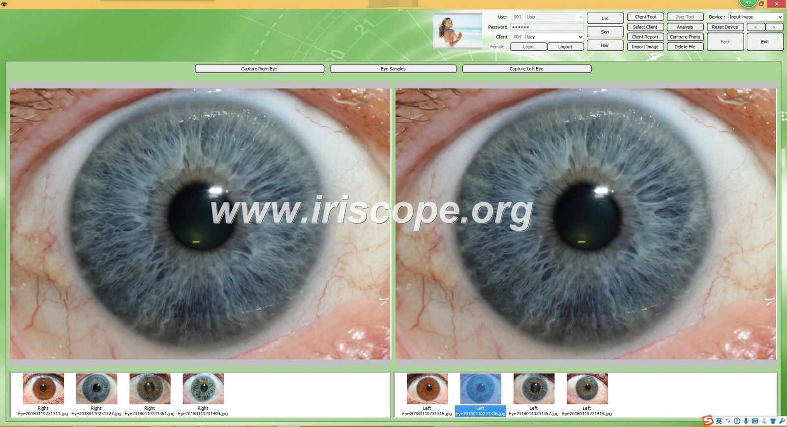
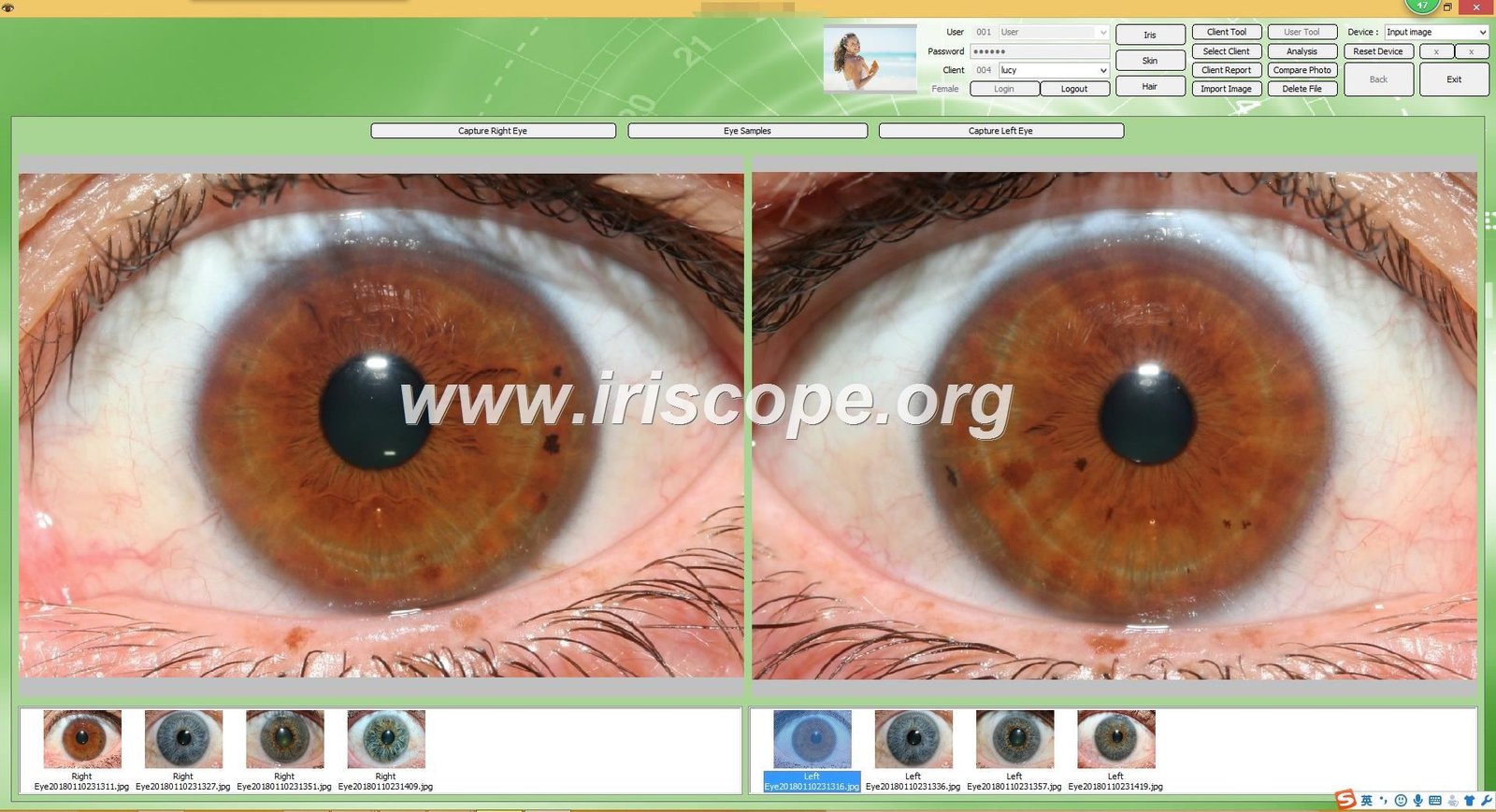
High-resolution iridology cameras capture detailed iris images under controlled lighting. These specialized cameras eliminate reflections while maximizing fiber visibility.
Professional lighting systems provide consistent, shadow-free illumination. Proper lighting reveals subtle color variations and depth patterns.
Magnification capabilities ranging from 20x to 50x enable thorough sector examination. Variable magnification helps identify both broad patterns and minute details.
Software Analysis Capabilities
Modern iridology software overlays anatomical charts directly onto captured iris images. This feature ensures precise sector identification.
Comparison tools display multiple images side-by-side for tracking changes. Sequential documentation reveals progressive patterns over months or years.
Measurement tools quantify lacunae size, pigment density, and fiber spacing. Objective measurements enhance interpretation consistency.
MAIKONG Professional Iridology Solutions
With over 20 years of research and manufacturing experience, MAIKONG produces professional-grade iridology equipment trusted by practitioners worldwide. Our comprehensive product line includes advanced iriscopes, high-resolution iridology cameras, detailed reference charts, and sophisticated analysis software.
MAIKONG offers the world’s first iridology software supporting both Windows (7/8/10/11) and Apple MAC OS operating systems. This cross-platform compatibility eliminates workflow barriers while maintaining professional-grade analytical capabilities.
Our direct factory pricing provides exceptional value without compromising quality. We support practitioners with multilingual software options, customizable branding through OEM/ODM services, and comprehensive technical training.
MAIKONG actively seeks qualified distributors and dealers throughout the United States. Partner with us to bring premium iridology tools to your market while building a profitable business relationship.
Core Product Advantages
- Direct factory pricing eliminates middleman costs
- Windows and MAC OS software compatibility
- Multilingual interface supporting global practice
- Comprehensive technical support via WhatsApp
- Custom branding and localization available
- Generous distributor profit margins
Our Product Range
- Professional iriscopes with LED illumination
- High-resolution iridology cameras (USB/wireless)
- Comprehensive iridology chart sets
- Advanced analytical software (Windows/MAC)
- Training materials and certification support
- Ongoing technical and business support
Documentation Best Practices
Maintain consistent photographic protocols for all client examinations. Use identical lighting, magnification, and positioning for comparable images.
Organize digital files systematically with clear naming conventions. Include date, client identifier, and eye designation in each filename.
Back up all client images securely in multiple locations. Professional practice standards require maintaining long-term records.
Integration with Comprehensive Health Assessment
Iridology provides valuable insights but functions best within broader health evaluation. Combining iris analysis with other assessment methods creates comprehensive understanding.
The left iris sectors examination complements rather than replaces conventional diagnostic approaches. Professional practitioners integrate multiple evaluation tools.
Holistic health assessment considers physical, emotional, and environmental factors. Iris patterns provide one piece of the larger wellness puzzle.
Correlating with Client History
Client interviews reveal lifestyle factors affecting respiratory health. Smoking history, occupational exposures, and exercise habits provide essential context.
Previous health conditions help interpret current iris findings. Childhood illnesses, past injuries, and chronic conditions leave lasting iris signatures.
Family health patterns indicate genetic predispositions visible in iris structure. Constitutional patterns pass through generations.
Supporting Wellness Strategies
Iris findings guide personalized wellness recommendations. Identified weaknesses suggest targeted support strategies.
Respiratory sector patterns inform breathing exercises, environmental modifications, and nutritional support. Specific interventions address observed vulnerabilities.
Follow-up examinations track intervention effectiveness over time. Sequential iris images document progressive improvements or persistent concerns.
Physical Assessment Tools
- Iris examination
- Tongue diagnosis
- Nail analysis
- Postural evaluation
- Vital sign measurement
Lifestyle Evaluation
- Dietary habits assessment
- Exercise patterns review
- Sleep quality evaluation
- Stress level analysis
- Environmental exposure history
Functional Testing
- Breath capacity measurement
- Oxygen saturation monitoring
- Peak flow testing
- Symptom tracking journals
- Response to interventions
Referral Relationships
Serious respiratory concerns identified through iris examination warrant medical referral. Professional practitioners maintain collaborative relationships with healthcare providers.
Communicating findings appropriately respects scope of practice boundaries. Iridology serves as assessment rather than diagnostic tool.
Integrated care teams provide comprehensive support for clients with complex health needs. Iris analysis contributes valuable information to collaborative treatment planning.
Advanced Interpretation Techniques
Experienced practitioners develop nuanced interpretation skills beyond basic pattern recognition. Advanced analysis considers subtle variations and complex combinations.
Mastery requires years of practice examining thousands of iris patterns. Pattern recognition abilities improve steadily with dedicated study and clinical experience.
The following advanced concepts enhance diagnostic precision and predictive capabilities. These techniques distinguish expert analysis from basic interpretation.
Temporal Analysis and Progression
Comparing sequential images reveals health trajectory over time. Progressive darkening suggests declining function while lightening indicates improvement.
New formations appearing in previously clear areas signal developing concerns. Early detection enables preventive intervention before symptoms manifest.
Stable patterns persisting across multiple examinations indicate constitutional characteristics. These permanent features guide long-term wellness strategies.
Bilateral Comparison Significance
Asymmetry between corresponding sectors in right and left eyes carries diagnostic weight. Significant differences warrant thorough investigation.
The left eye lung sector compared with the right eye corresponding zone reveals bilateral lung health differences. Unilateral patterns may indicate localized issues.
Symmetrical patterns suggest system-wide influences affecting both lungs equally. Constitutional factors typically create bilateral symmetry.
Pupillary Response Assessment
The pupil position and shape provide additional diagnostic clues. Displacement toward specific sectors indicates nerve stress in corresponding organs.
A pupil pulling toward the lung sector suggests sympathetic nervous system activation. This pattern correlates with respiratory stress responses.
Irregular pupil borders near the lung zone show localized nerve irritation. These subtle distortions require careful observation to detect.


Case Study Applications
Real-world examples demonstrate practical application of left iris sectors interpretation. These cases illustrate how theoretical knowledge translates to clinical practice.
Client confidentiality protections require anonymizing identifying details. The following composite cases represent typical presentation patterns.
Each case demonstrates the integration of iris findings with client history and wellness outcomes. These examples guide similar situation interpretation.
Case Example: Chronic Respiratory Weakness
A 42-year-old client presented with frequent respiratory infections. Iris examination revealed multiple small lacunae in the right lung sector.
Deep stress rings crossed through the respiratory zone bilaterally. Orange pigment deposits indicated historical toxic burden.
Client history confirmed childhood asthma and 15-year smoking history ended three years prior. The iris patterns correlated perfectly with reported health challenges.
Wellness recommendations focused on respiratory strengthening exercises and immune support. Follow-up examination six months later showed slightly improved fiber density.
Case Example: Acute Inflammatory Response
A 28-year-old client reported recent onset of persistent cough. Iris analysis showed bright white fiber elevation in the upper right lung sector.
The acute inflammatory pattern appeared sharply defined with surrounding redness. No other significant constitutional weakness patterns were present.
Medical evaluation confirmed bronchitis requiring treatment. The iris sign resolved completely after infection cleared three weeks later.
This case demonstrated iris analysis detecting acute processes distinct from constitutional patterns. Appropriate medical referral ensured proper care.
Case Example: Constitutional Predisposition
A 35-year-old client with no current respiratory complaints sought preventive wellness guidance. Large bilateral lacunae appeared in both lung sectors.
Family history revealed multiple relatives with chronic obstructive pulmonary disease. The iris pattern indicated genetic predisposition.
Preventive recommendations included avoiding respiratory irritants and maintaining optimal lung capacity through regular cardiovascular exercise. Annual monitoring tracks any progressive changes.
Limitations and Realistic Expectations
Iridology offers valuable insights but has important limitations requiring acknowledgment. Understanding these boundaries ensures appropriate practice and ethical client communication.
Professional practitioners clearly explain what iris analysis can and cannot determine. Setting realistic expectations builds trust and prevents misunderstanding.
The following limitations apply to iris examination across all applications. Recognizing these constraints improves practice quality and professional credibility.
What Iris Analysis Cannot Do
Iridology cannot diagnose diseases with medical certainty. Iris patterns indicate tendencies and predispositions rather than confirmed conditions.
Iris examination cannot determine specific pathogen identity. Whether bacterial, viral, or fungal infections cause observed patterns requires laboratory testing.
Iris analysis cannot predict exact disease onset timing. Constitutional weakness may never manifest as clinical illness depending on lifestyle factors.
Scope of Practice Considerations
Professional boundaries vary by jurisdiction and practitioner credentials. Many regions restrict diagnosing diseases to licensed medical providers.
Iridologists should clearly communicate their role as wellness consultants rather than medical diagnosticians. Appropriate language acknowledges assessment versus diagnosis.
Serious health concerns identified through iris examination require medical referral. Professional responsibility includes recognizing when conventional care becomes necessary.
Important Professional Note: Iridology serves as a complementary assessment tool within holistic health practice. It does not replace conventional medical diagnosis or treatment. Practitioners should maintain appropriate referral relationships with licensed healthcare providers and clearly communicate the scope and limitations of iris analysis to all clients.
Research and Evidence Considerations
Scientific research on iridology shows mixed results with methodological challenges. Standardization difficulties complicate controlled study design.
Clinical observation over decades provides substantial empirical evidence. Many practitioners report consistent correlations between iris patterns and client health conditions.
Ongoing research continues exploring iris–body relationships using modern imaging technology. Future studies may clarify underlying mechanisms and improve interpretation precision.
Continuing Education and Skill Development
Mastering left iris sectors interpretation requires ongoing learning beyond initial training. Professional development enhances accuracy and expands capabilities.
Multiple pathways support practitioner growth from beginner through advanced expert levels. Dedication to continuous improvement distinguishes exceptional practitioners.
The following resources and strategies accelerate skill development in iris analysis. Combining formal education with practical experience yields optimal results.
Certification Programs and Training
Formal iridology certification programs provide structured foundational education. These courses cover anatomy, chart interpretation, and professional practice standards.
Certification requirements typically include classroom hours, practical examinations, and supervised case studies. Credentials demonstrate commitment to professional standards.
Advanced specialty certifications focus on specific interpretation techniques or client populations. Continuing education maintains certification status in most programs.
Mentorship and Peer Learning
Studying under experienced practitioners accelerates skill acquisition. Mentors provide real-time feedback on interpretation accuracy and technique refinement.
Case review groups allow collaborative learning from diverse clinical experiences. Group discussion reveals multiple interpretation perspectives.
Professional associations facilitate networking and knowledge exchange. Conference attendance exposes practitioners to cutting-edge developments and techniques.
Building Pattern Recognition Skills
Examining hundreds of iris images develops pattern recognition abilities. Regular practice with diverse constitutional types improves diagnostic precision.
Maintaining personal iris image libraries organized by pattern type creates reference resources. Reviewing these collections reinforces learning and reveals subtle variations.
Tracking client outcomes over time validates interpretation accuracy. Correlation between iris findings and subsequent health developments confirms or refines understanding.
Explore Professional Development Resources
MAIKONG supports practitioner success through comprehensive training materials and ongoing educational resources. Our commitment extends beyond equipment sales to fostering professional excellence throughout the iridology community.
We provide access to certification program information, reference materials, and technical training for our equipment users. Partner practitioners receive priority support and business development assistance.
Frequently Asked Questions
How accurate is left iris sectors interpretation for lung health assessment?
Accuracy depends heavily on practitioner experience and proper equipment use. Experienced practitioners report strong correlations between iris patterns and client respiratory history.
Iris analysis identifies constitutional tendencies and potential vulnerabilities rather than confirming specific diseases. It functions best as part of comprehensive health assessment rather than standalone diagnostic tool.
Professional iridology equipment significantly improves observation precision. High-resolution imaging reveals subtle patterns invisible during basic visual examination. For technical guidance on optimal equipment selection, contact experienced practitioners or specialized equipment manufacturers.
Can iris patterns change over time with improved health?
Some iris patterns can shift with significant health improvements while others remain relatively stable. Acute inflammatory signs like white fiber elevation typically resolve as conditions heal.
Toxic deposits may lighten gradually with detoxification and improved elimination. Fiber density can improve modestly with enhanced nutrition and reduced stress.
Constitutional patterns like large lacunae generally persist throughout life as they represent inherent tissue structure. However, supporting overall health can prevent these predispositions from manifesting as clinical conditions.
Sequential iris photography over months or years documents these changes. Professional tracking requires consistent imaging protocols and quality equipment.
What is the difference between the left eye iridology chart and right eye chart?
The left eye iridology chart maps the right side of your body while the right eye chart corresponds to your left body structures. This mirror relationship applies consistently across all body systems.
Your right lung appears in the left iris at approximately 2-3 o’clock. Your left lung shows in the right iris at the corresponding 9-10 o’clock position.
Both charts follow identical organizational principles with organs and systems arranged in specific sectors. The digestive system surrounds the pupil in both eyes, while extremities appear in the peripheral zones.
Professional practitioners examine both eyes to assess bilateral body symmetry and identify side-specific concerns. Comprehensive analysis requires comparing corresponding sectors across both eyes.
Do I need special equipment to practice iridology professionally?
Professional practice requires at minimum a quality magnification tool and proper lighting. Basic examination is possible with a strong magnifying glass and penlight, though this limits diagnostic precision.
Serious practitioners invest in dedicated iridology cameras providing magnification, controlled lighting, and image capture capabilities. Documentation through photography enables tracking changes over time and comparing with reference images.
Professional software enhances interpretation accuracy by overlaying anatomical charts and providing measurement tools. Modern systems supporting both Windows and MAC OS eliminate technology barriers.
Equipment quality directly impacts diagnostic capabilities and professional credibility. Clients expect professional tools during consultations. For equipment recommendations matching your practice needs and budget, consult with established practitioners or contact manufacturers directly.
How long does it take to become proficient at iris interpretation?
Basic competency typically requires 6-12 months of structured study and practice. Initial training covers fundamental anatomy, chart orientation, and major pattern recognition.
Intermediate proficiency develops over 2-3 years with consistent practice examining diverse clients. Pattern recognition skills improve steadily through repeated observation and outcome tracking.
Advanced expertise requires 5-10 years of dedicated practice examining thousands of iris patterns. Master practitioners develop intuitive recognition of subtle variations and complex pattern combinations.
Formal certification programs typically require 100-200 training hours plus supervised case studies. Continuing education maintains and expands skills throughout professional careers.
Mentorship under experienced practitioners significantly accelerates skill development compared to self-study alone.
Can iridology detect lung cancer or other serious diseases?
Iridology cannot diagnose diseases like cancer with medical certainty. Iris patterns reveal constitutional tendencies, tissue weakness, and system stress rather than specific pathological conditions.
Unusual or concerning patterns warrant medical referral for conventional diagnostic testing. Iris analysis may identify areas requiring closer attention but cannot replace medical imaging, biopsy, or laboratory testing.
Some practitioners report observing iris changes preceding clinical diagnosis of serious conditions. However, these observations lack sufficient research validation for definitive diagnostic claims.
Professional responsibility requires clearly communicating iridology limitations and maintaining appropriate referral relationships. Iris analysis serves best as complementary assessment within integrated health care approaches.
Clients presenting with serious symptoms require immediate medical evaluation regardless of iris findings.
What training or certification do I need to practice iridology?
Requirements vary significantly by location as iridology regulation differs across jurisdictions. Some regions have no specific iridology licensing requirements while others include it under natural medicine or complementary therapy regulations.
Professional associations offer voluntary certification programs establishing practice standards. These credentials demonstrate commitment to professional excellence even where not legally required.
Many iridologists hold primary credentials in related fields like naturopathy, nutrition, or holistic health. Iridology training then enhances their existing practice scope.
Comprehensive training programs cover anatomy, physiology, pattern interpretation, professional ethics, and business practices. Quality programs include supervised practical experience alongside theoretical study.
Ongoing continuing education maintains and expands skills throughout professional careers. For specific regional requirements and recognized training programs, consult local professional associations or regulatory bodies.
How do environmental factors appear in the left iris sectors?
Environmental exposures create characteristic pigment deposits and tissue changes in corresponding iris sectors. Toxic burden accumulation often indicates through orange, yellow, or dark brown discoloration.
Smoking history typically produces distinctive orange-rust deposits in lung sectors. These patterns may persist years after smoking cessation, reflecting long-term tissue impact.
Occupational exposures to dust, chemicals, or pollutants create similar pigment accumulations. The specific coloration and pattern sometimes suggests exposure type.
Air quality in living environments influences respiratory sector appearance over time. Urban versus rural residence histories may correlate with observable iris differences.
Supporting natural detoxification processes can gradually lighten toxic deposits. However, deeply encapsulated old exposures may remain visible permanently.
Conclusion
Mastering left iris sectors interpretation for right lung assessment requires dedicated study, quality equipment, and extensive practice. The techniques outlined throughout this guide provide a comprehensive foundation for professional application.
Your iris reveals remarkable information about constitutional strengths and vulnerabilities. The right lung sector specifically offers valuable insights into respiratory health patterns and predispositions.
Professional practice demands ongoing skill development and appropriate scope of practice boundaries. Iridology functions optimally as part of comprehensive health assessment rather than standalone diagnostic method.
Quality equipment investments directly impact diagnostic capabilities and professional credibility. Modern technology has significantly enhanced iris examination precision and documentation abilities.
Whether you’re beginning your iridology journey or refining advanced skills, consistent practice and continuing education remain essential. The field continues evolving as research and clinical experience expand our understanding.
Professional practitioners combine technical expertise with compassionate client care. Clear communication about iridology capabilities and limitations builds trust and maintains ethical standards.
Your commitment to excellence in iris analysis serves your clients’ wellness goals while advancing the professional field. Each examination contributes to the growing body of clinical observations and outcomes.