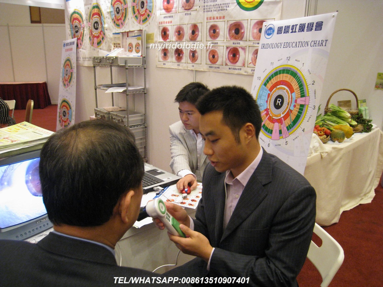
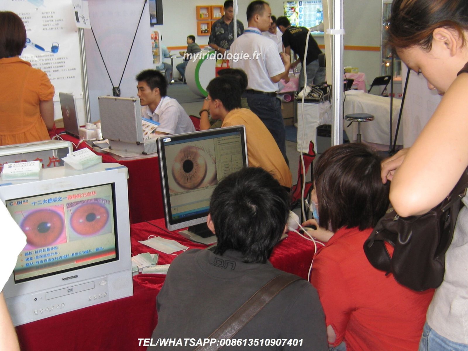
The human iris contains over 28,000 nerve fibers connected to every tissue and organ in the body, making it a potential window into our overall health. Professional иридология, диагностика радужной оболочки глаза examinations analyze these intricate patterns to identify potential health imbalances before they manifest as physical symptoms. This comprehensive guide explores how qualified practitioners conduct these assessments, the tools they use, and the insights they can provide about your wellbeing.

Профессиональный иридолог осматривает радужную оболочку пациента с помощью специального оборудования.
Iridology iris diagnosis is a holistic assessment method that examines the colored part of the eye to evaluate overall health and potential areas of concern in the body. Dating back to the 19th century, this practice was pioneered by Hungarian physician Dr. Ignatz von Peczely, who first observed correlations between iris markings and physical conditions.
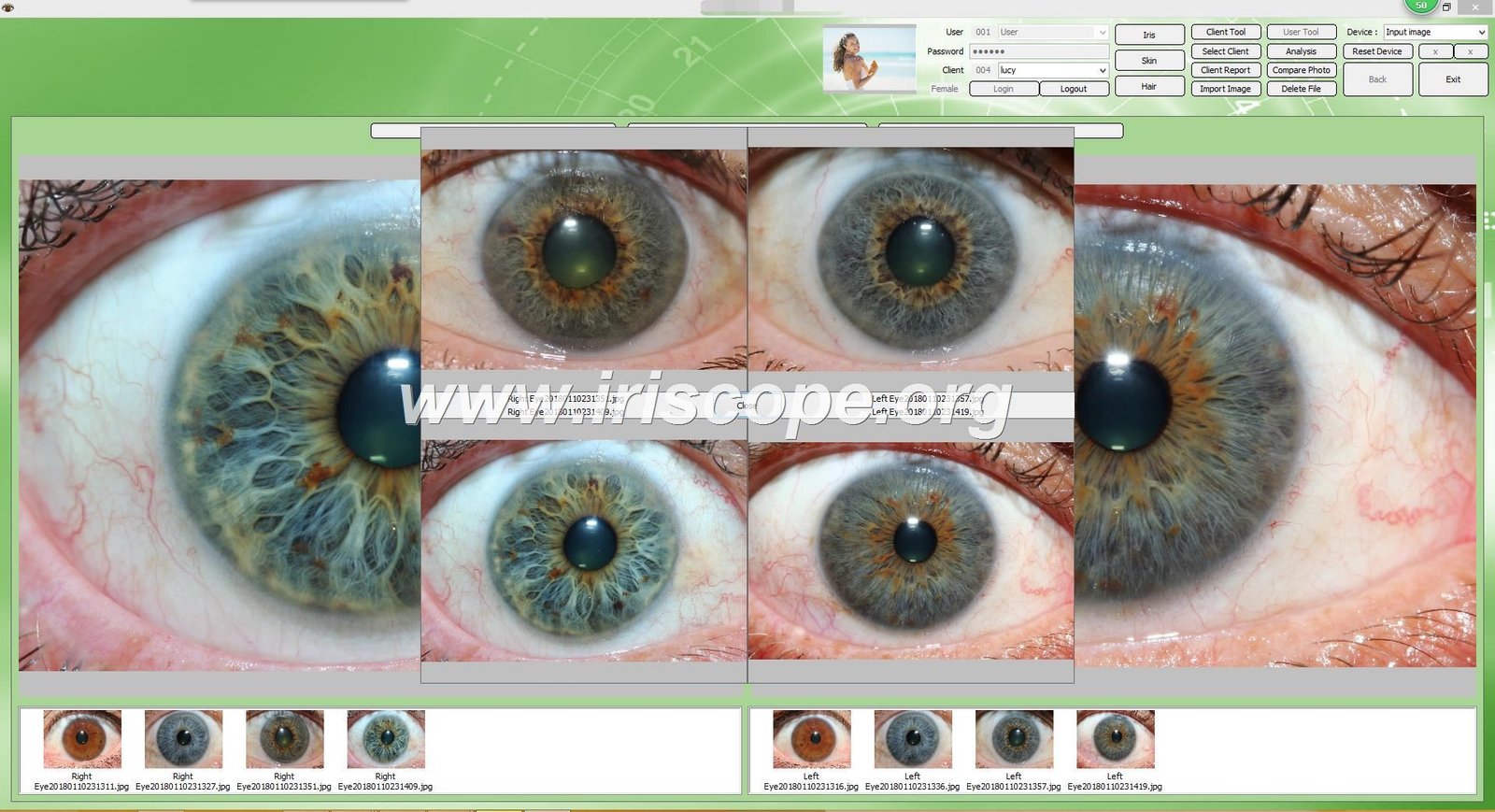
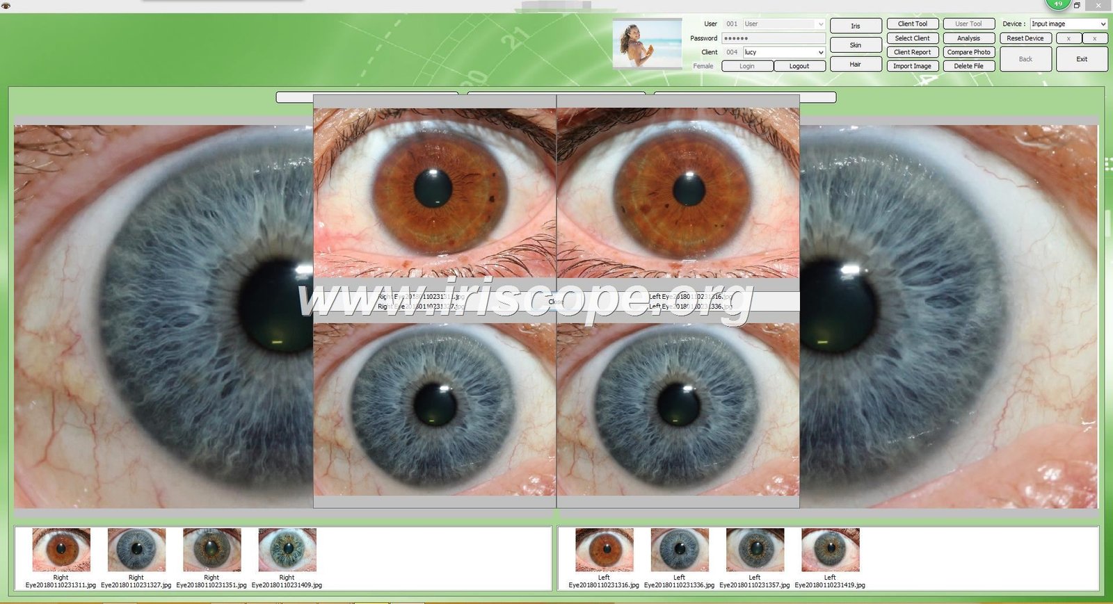
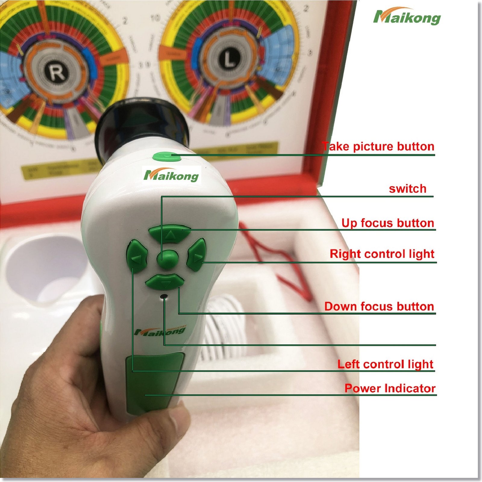
Modern iridology has evolved significantly since its inception, incorporating advanced imaging technology and standardized analysis methods. Today’s practitioners use digital iriscopes and sophisticated software to capture high-resolution images of the iris and identify subtle patterns that may indicate health imbalances.
While conventional medicine approaches illness after symptoms appear, иридология, диагностика радужной оболочки глаза aims to identify potential weaknesses before they manifest as disease. This preventative approach aligns with growing interest in proactive health management and complementary assessment methods.
What is Иридология Диагностика радужной оболочки?

Iridology chart mapping iris zones to corresponding body systems
Iridology iris diagnosis is based on the principle that the iris reflects the condition of tissues throughout the body through neurological connections. Each area of the iris corresponds to specific organs and systems according to standardized iridology charts. By examining the color, structure, and markings in these areas, practitioners can identify potential health issues.
The science behind iridology centers on the concept that the iris contains nerve endings connected to the brain via the optic nerve. These connections form a complex communication network with all body tissues. When an organ or system experiences stress or dysfunction, these changes may be reflected in the corresponding iris zone.
Key Principles of Иридология Диагностика радужной оболочки
- Constitutional Assessment: Evaluating inherent strengths and weaknesses in body systems
- Inflammation Markers: Identifying areas of acute or chronic inflammation
- Toxicity Indicators: Recognizing signs of toxic accumulation
- Nutritional Status: Assessing potential nutritional deficiencies
- Stress Response: Evaluating how the body responds to physical and emotional stress
It’s important to note that while iridology has passionate advocates, the scientific community remains divided on its efficacy. Some studies question its diagnostic reliability, while practitioners point to clinical success and patient testimonials. Iridology iris diagnosis is best viewed as a complementary assessment tool rather than a replacement for conventional medical diagnosis.
Steps in a Professional Иридология Диагностика радужной оболочки Exam


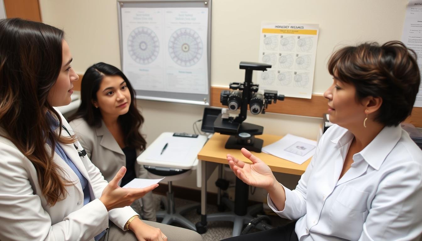
Iridologist conducting a thorough pre-examination consultation
Профессионал иридология, диагностика радужной оболочки глаза examination follows a structured approach to ensure comprehensive assessment and accurate interpretation. The following steps outline the typical process used by qualified practitioners.
Pre-Exam Consultation
Before examining the iris, a professional iridologist conducts a detailed consultation to gather essential background information. This typically includes:
- Comprehensive health history review
- Current symptoms and concerns
- История здоровья семьи
- Факторы образа жизни (диета, физические упражнения, уровень стресса)
- Современные лекарства и добавки
This information provides crucial context for interpreting iris signs and helps the practitioner develop a more personalized assessment. The consultation also establishes rapport and helps the client understand what to expect during the examination.
Iris Imaging Techniques

Digital iriscope capturing high-resolution iris images
Capturing clear, detailed images of the iris is essential for accurate analysis. Modern practitioners use specialized equipment including:
- Digital iriscopes with high-resolution cameras
- Magnification systems (typically 10-30x)
- Specialized lighting to reveal subtle iris details
- Image storage software for comparison over time
The imaging process is non-invasive and comfortable for the client. The practitioner will typically capture images of both the left and right irises, as well as close-up sections of specific areas of interest. These images are then displayed on a screen for immediate analysis or saved for more detailed examination.
Analysis of Iris Markings

Detailed analysis of iris markings using specialized software
The core of иридология, диагностика радужной оболочки глаза lies in the systematic analysis of iris characteristics. Practitioners evaluate:
- Iris color and pigmentation variations
- Structural integrity of iris fibers
- Presence of specific markings (lacunae, crypts, radii solaris)
- Pupil size, shape, and reactivity
- Sclera (white of the eye) coloration and markings
Using standardized iridology charts, the practitioner maps these observations to corresponding body systems. Advanced software may assist in identifying patterns and providing reference comparisons. The analysis considers both isolated markings and the overall iris constitution.
Diagnostic Reporting

Iridologist explaining diagnostic findings using detailed iris images
After completing the analysis, the practitioner prepares a comprehensive report of their findings. This typically includes:
- Overview of constitutional strengths and weaknesses
- Identification of potential areas of concern
- Correlation with symptoms and health history
- Personalized recommendations for health optimization
- Visual documentation with annotated iris images
The practitioner reviews these findings with the client, explaining the significance of various iris signs and answering questions. This educational component helps clients understand the connections between iris markings and their health status.
Ready to experience professional iridology?
Our team of experts provides comprehensive иридология, диагностика радужной оболочки глаза using state-of-the-art equipment. Contact us today to schedule your consultation.
Связаться через WhatsApp
Call: +86-13-51090-74-01
Case Studies: Real-World Applications of Иридология Диагностика радужной оболочки

Iridologist reviewing comprehensive case findings with a patient
The following case studies illustrate how professional иридология, диагностика радужной оболочки глаза has been applied in real-world scenarios. These examples demonstrate the potential insights and benefits of this assessment method while acknowledging its complementary role alongside conventional healthcare.
Case Study 1: Identifying Digestive System Weaknesses

Iris showing distinctive markings in the digestive system zone
Patient Profile: 42-year-old female with chronic fatigue, bloating, and intermittent digestive discomfort
Ирис Наблюдения: The iridology examination revealed significant lacunae (hollow areas) in the zones corresponding to the small intestine and pancreas. The practitioner also noted a darkened area in the liver zone of the right iris.
Корреляция: These iris signs aligned with the patient’s reported digestive symptoms. The practitioner suggested potential issues with nutrient absorption and liver function.
Outcome: Based on these findings, the patient consulted with her physician who ordered additional testing. Lab work confirmed mild pancreatic enzyme insufficiency and elevated liver enzymes. A targeted treatment plan addressing these specific issues led to significant symptom improvement within three months.
Case Study 2: Constitutional Stress Patterns

Iris displaying characteristic nervous system stress patterns
Patient Profile: 35-year-old male with recurring headaches, sleep disturbances, and anxiety
Ирис Наблюдения: Iridology assessment revealed pronounced nerve rings (circular lines) in both irises and a concentration of stress fibers in the cerebral zone. The overall iris structure showed a neurogenic constitution type.
Корреляция: These patterns indicated constitutional sensitivity to stress and potential nervous system overactivity, aligning with the patient’s symptoms.
Outcome: The practitioner recommended targeted stress management techniques, specific nutritional support for the nervous system, and lifestyle modifications. After implementing these recommendations and working with his healthcare provider, the patient reported a 70% reduction in headache frequency and improved sleep quality.
Case Study 3: Identifying Lymphatic Congestion

Iris showing indicators of lymphatic system congestion
Patient Profile: 51-year-old female with chronic sinus congestion, frequent minor infections, and fatigue
Ирис Наблюдения: The iridology examination revealed a pronounced lymphatic rosary (a ring of discoloration in the outer iris) and cloudy areas in the lymphatic zones. The practitioner also noted tophi (small white spots) scattered throughout both irises.
Корреляция: These signs suggested potential lymphatic congestion and accumulation of metabolic waste products, consistent with the patient’s recurring infections and fatigue.
Outcome: Based on these findings, the patient implemented a comprehensive lymphatic support program including hydration, specific movement practices, and botanical support. Within two months, she reported improved energy levels and reduced frequency of infections. Her conventional healthcare provider noted improved lymphatic drainage upon physical examination.
These case studies illustrate how иридология, диагностика радужной оболочки глаза can provide valuable insights when used as part of a comprehensive health assessment. While not a replacement for conventional medical diagnosis, iridology can help identify potential areas of weakness that may benefit from targeted support and further investigation.
Часто задаваемые вопросы О нас Иридология Диагностика радужной оболочки

Professional iridologist addressing common questions about the examination process
Является Иридология Диагностика радужной оболочки scientifically validated?
The scientific status of iridology remains contested. While some studies have questioned its diagnostic reliability, others have shown correlations between iris signs and certain health conditions. Iridology is best viewed as a complementary assessment tool rather than a definitive diagnostic method. It can provide insights into constitutional tendencies and potential areas of weakness, but should be used alongside conventional medical evaluation.
How long does a professional iridology examination take?
A comprehensive иридология, диагностика радужной оболочки глаза session typically takes 60-90 minutes. This includes the initial consultation (15-20 minutes), iris imaging (10-15 minutes), analysis (20-30 minutes), and review of findings with recommendations (15-25 minutes). Follow-up sessions are usually shorter, focusing on changes and progress.
Can iridology detect specific diseases?
Iridology is not designed to diagnose specific diseases. Rather, it identifies potential areas of weakness, inflammation, or congestion in body systems. These insights can help direct attention to areas that may benefit from support or further medical investigation. Ethical practitioners emphasize that iridology complements rather than replaces conventional medical diagnosis.
How often should I have an iridology examination?
For general health monitoring, annual examinations are typically sufficient. Those implementing specific health protocols based on iridology findings might benefit from follow-up assessments every 3-6 months to track changes. The appropriate frequency depends on individual health goals and circumstances.
What qualifications should I look for in an iridologist?
Look for practitioners with formal training from recognized iridology institutions, certification from professional organizations, and continuing education in the field. Many qualified iridologists also have backgrounds in healthcare professions such as naturopathy, chiropractic, or nursing. Ask about their experience, approach, and whether they work collaboratively with other healthcare providers.
Can children undergo iridology examinations?
Yes, iridology is suitable for all ages, including children. The non-invasive nature of the examination makes it particularly appropriate for pediatric assessment. Children’s irises can provide insights into constitutional tendencies and potential health patterns. The examination process is typically modified to accommodate shorter attention spans and comfort levels.
Will wearing contact lenses affect my iridology examination?
Contact lenses should be removed before an iridology examination as they can obscure important iris details and potentially distort findings. If you wear contacts, bring your lens case and solution to the appointment. Those who wear colored contacts should remove them at least 24 hours before the examination to allow the iris to return to its natural state.
How do iris signs change over time?
While the basic constitutional pattern of the iris remains relatively stable throughout life, certain iris signs can change in response to health developments. Acute conditions may be reflected in temporary changes, while chronic issues may lead to more persistent markings. Regular iridology examinations can help track these changes and provide insights into the effectiveness of health interventions.
Can iridology help with preventative health?
Preventative health is one of the strongest applications of iridology. By identifying constitutional weaknesses and potential problem areas before symptoms develop, iridology can guide targeted preventative strategies. This proactive approach aligns with the growing emphasis on preventative healthcare and personalized wellness protocols.
How does iridology differ between the left and right eyes?
In traditional iridology, the left iris generally corresponds to the left side of the body and is associated more with the lymphatic system, while the right iris corresponds to the right side and relates more to the hepatic (liver) system. Some schools of iridology also associate the left eye with feminine/receptive energy and the right with masculine/projective energy. Both irises are examined for a complete assessment.
Заключение
Профессионал иридология, диагностика радужной оболочки глаза offers a unique perspective on health assessment by examining the intricate patterns of the iris. While not a replacement for conventional medical diagnosis, this non-invasive approach can provide valuable insights into constitutional tendencies, potential areas of weakness, and overall health patterns.
The effectiveness of iridology depends significantly on the practitioner’s training, experience, and the quality of equipment used. When performed by qualified professionals using modern tools and standardized methods, иридология, диагностика радужной оболочки глаза can contribute meaningful information to a comprehensive health assessment.
Whether you’re a health practitioner interested in adding iridology to your skill set or an individual curious about what your eyes might reveal about your health, understanding the professional approach to iridology helps ensure you receive the most accurate and beneficial assessment possible.
Experience Professional Iridology Services
Our team provides comprehensive иридология, диагностика радужной оболочки глаза using state-of-the-art equipment. We also offer professional iridology tools for practitioners.
Call: +86-13-51090-74-01

