Maikong-Professional Iridology Camera Iriscope Výrobca a dodávateľ

1>Iris Medical Term: Anatomy, Functions, and Related Conditions

» The Ultimate Guide to Iris Eye Care: Protecting Your Vision Naturally » Iris Medical Term: Anatomy, Functions, and Related Conditions

Iris Medical Term: The iris medical term refers to the colored part of your eye that surrounds the pupil. This remarkable structure does far more than give your eyes their distinctive color—it plays a crucial role in vision by controlling how much light enters your eye. Understanding the iris’s anatomy and functions can help you recognize potential eye problems early and maintain optimal eye health.
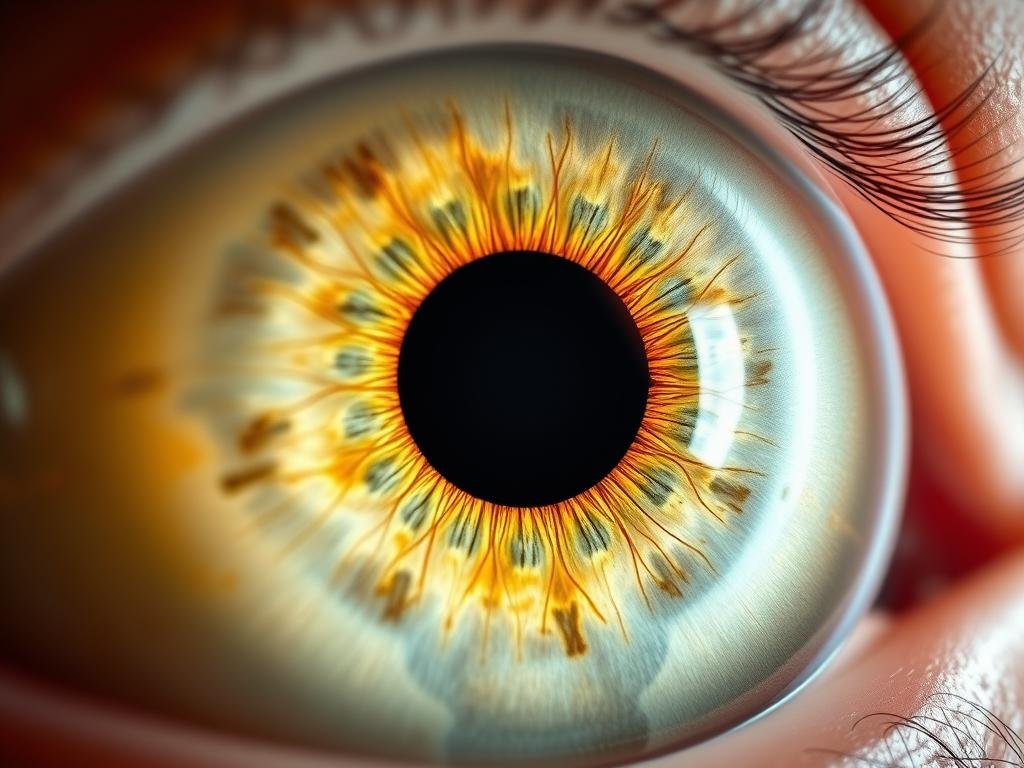
Close-up of a human iris medical term showing detailed structure

The iris is the colored part of the eye that controls pupil size

What is the Iris? Medical Term Definition

The iris is the opaque, pigmented continuation of the choroid layer of the eye. It partially covers the lens, surrounding the pupil and regulating the amount of light that strikes the retina. Located between the cornea and lens, the iris divides the anterior chamber from the posterior chamber of the aqueous compartment.

Much like your fingerprint, the color and pattern of your iris is unique to you. No other person in the world has exactly the same iris structure or coloration, which is why iris scanning has become a reliable biometric identification method.

Need Expert Eye Care?

Regular eye exams can detect potential issues before they affect your vision.

Schedule an Eye Exam Today

Iris Anatomy and Structure

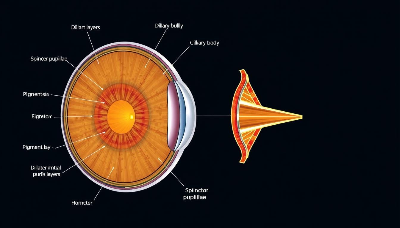
Anatomical diagram of the iris medical term showing muscles and layers

Cross-section of the iris showing its muscular structure

The iris consists of several key components that work together to control pupil size and give the eye its color:

Muscular Structure

The iris contains two sets of muscles that work in opposition to control pupil size:

  • Sphincter pupillae muscleCircular muscles that contract to constrict the pupil in bright light (miosis)
  • Dilator pupillae muscleRadial muscles that contract to widen the pupil in dim light (mydriasis)

These muscles in the iris respond automatically to light conditions without conscious control, working through the parasympathetic and sympathetic nervous systems.

Pigmentation and Color

The color of your iris depends on the concentration of melanocytes (pigment-producing cells) in the iris stroma:

  • Low melanin concentration creates blue eyes
  • Medium melanin concentration creates green or hazel eyes
  • High melanin concentration creates brown eyes

Eye color is primarily determined by genetics, with specific genes controlling melanin production in the iris. The genes that determine your eye color are inherited from your biological parents.

Connection to Other Eye Structures

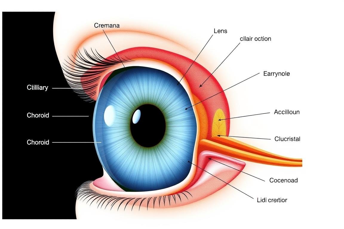
The iris is connected to the ciliary body, which contains the ciliary muscles that control the shape of the lens for focusing. Together with the choroid, these structures form the uveal tract, the middle layer of the eye between the sclera (outer layer) and retina (inner layer).

Diagram showing the iris medical term in relation to other eye structures

The iris in relation to other eye structures

Key Functions of the Iris

Light Regulation

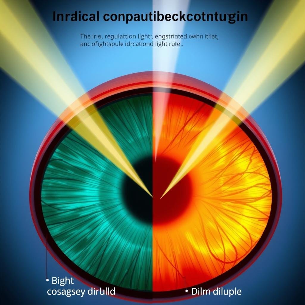
Illustration of iris medical term regulating light through pupil constriction and dilation

The primary function of the iris is to control the amount of light that enters the eye by adjusting pupil size. In bright environments, the sphincter muscles contract to make the pupil smaller, reducing light entry. In dim conditions, the dilator muscles contract to make the pupil larger, allowing more light to reach the retina.

UV Protection

Diagram showing how the iris medical term helps protect the eye from UV damage

The melanin pigment in the iris helps absorb ultraviolet (UV) radiation, providing some natural protection against UV damage to the internal structures of the eye. People with darker irises typically have greater natural UV protection than those with lighter colored eyes.

Aesthetic Role

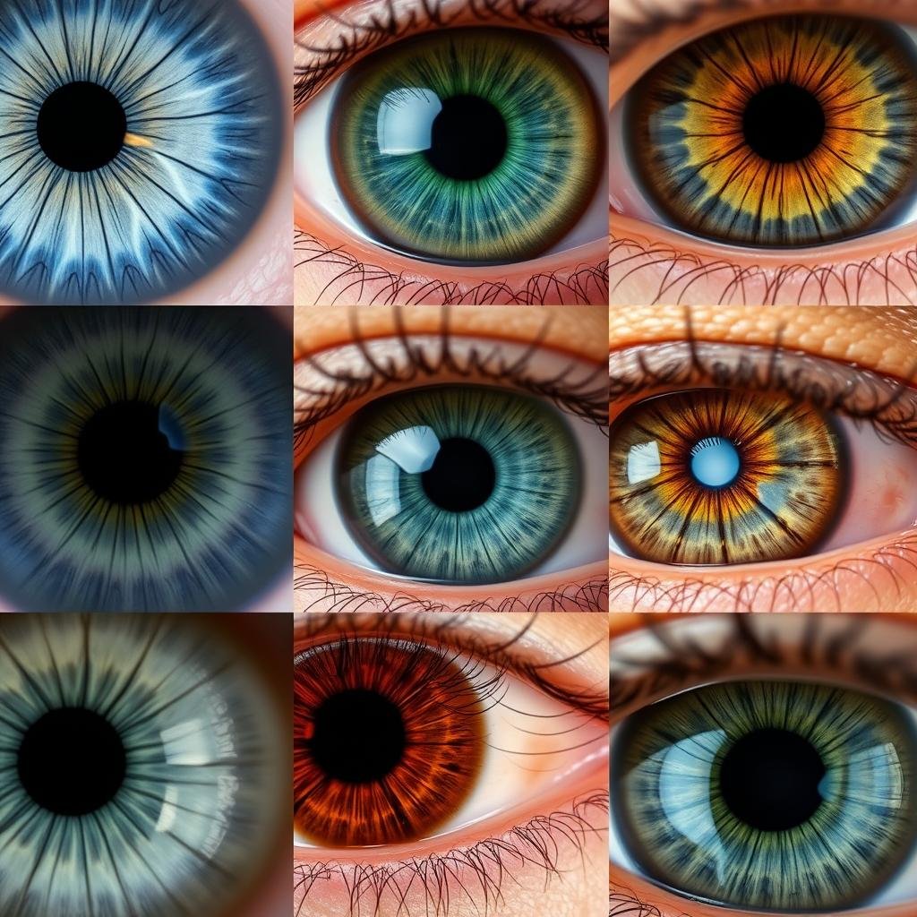
Close-up images of different colored irises showing unique patterns

Beyond its physiological functions, the iris gives each person their unique eye color and pattern. The distinctive appearance of each iris is determined by genetic factors and develops during early childhood, with some babies experiencing color changes in their first year of life.

The iris works constantly throughout your life, automatically adjusting to changing light conditions thousands of times each day to optimize your vision.

American Academy of Ophthalmology

Common Conditions Affecting the Iris Medical Term

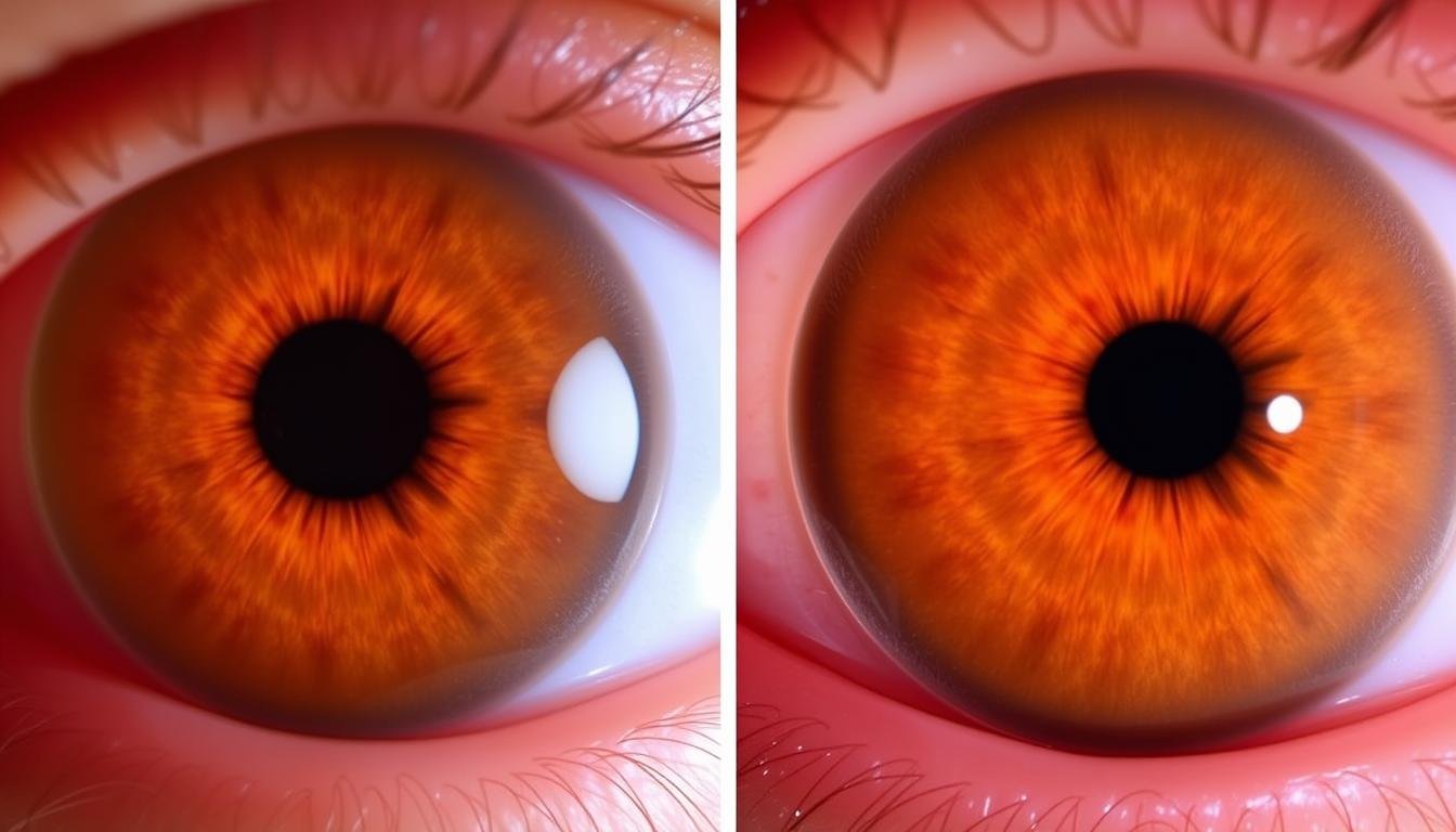
Comparison of healthy iris medical term versus one with iritis showing inflammation

Comparison between a healthy iris (left) and one with iritis (right)

Several conditions can affect the iris, potentially impacting vision and eye health:

Stav Opis Príznaky Treatment Approaches
Iritis (Anterior Uveitis) Inflammation of the iris, often associated with autoimmune disorders Eye pain, light sensitivity, blurred vision, redness Steroid eye drops, dilating drops, treating underlying causes
Aniridia Congenital condition where the iris is partially or completely absent Light sensitivity, poor vision, nystagmus (involuntary eye movements) Tinted contact lenses, sunglasses, management of associated conditions
Coloboma Gap or notch in the iris due to incomplete formation during development Keyhole-shaped pupil, light sensitivity, vision issues Tinted contact lenses, sunglasses, vision correction
Heterochromia Different colored irises or sections within one iris Usually asymptomatic, may indicate underlying conditions if acquired later in life None needed if congenital; treatment of underlying cause if acquired
Iris Melanoma Rare cancer that develops from pigment cells in the iris Growing dark spot on iris, change in pupil shape, vision changes Radiation therapy, surgical removal, monitoring

Warning: Seek immediate medical attention if you experience sudden eye pain, vision changes, light sensitivity, or notice changes in your iris appearance. These could indicate serious conditions requiring prompt treatment.

Experiencing Eye Symptoms?

Don’t wait until vision problems worsen. Consult with an ophthalmologist to address potential iris conditions early.

Find an Ophthalmologist Near You

Diagnostic Methods for Iris Examination

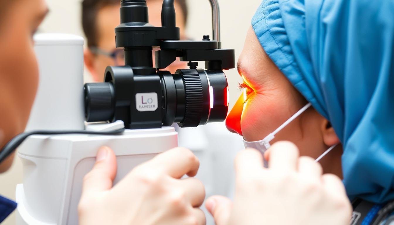
Doctor performing slit-lamp examination of patient's iris medical term

Slit-lamp examination allows detailed inspection of the iris

Eye care professionals use several methods to examine the iris and diagnose related conditions:

Slit-Lamp Examination

The slit lamp is a microscope with a focused light source that allows the ophthalmologist to examine the iris in great detail. This examination can reveal:

  • Inflammation or abnormal blood vessels
  • Structural abnormalities or damage
  • Deposits, nodules, or unusual pigmentation
  • Changes in iris texture or pattern

Pupillary Reflex Tests

These tests evaluate how well the iris muscles respond to light stimuli:

  • Direct pupillary reflexPupil constriction when light is shined into that eye
  • Consensual pupillary reflexPupil constriction in one eye when light is shined into the other eye
  • Accommodation reflexPupil constriction when focusing on a near object

Advanced Imaging Techniques

When more detailed visualization is needed, specialists may use:

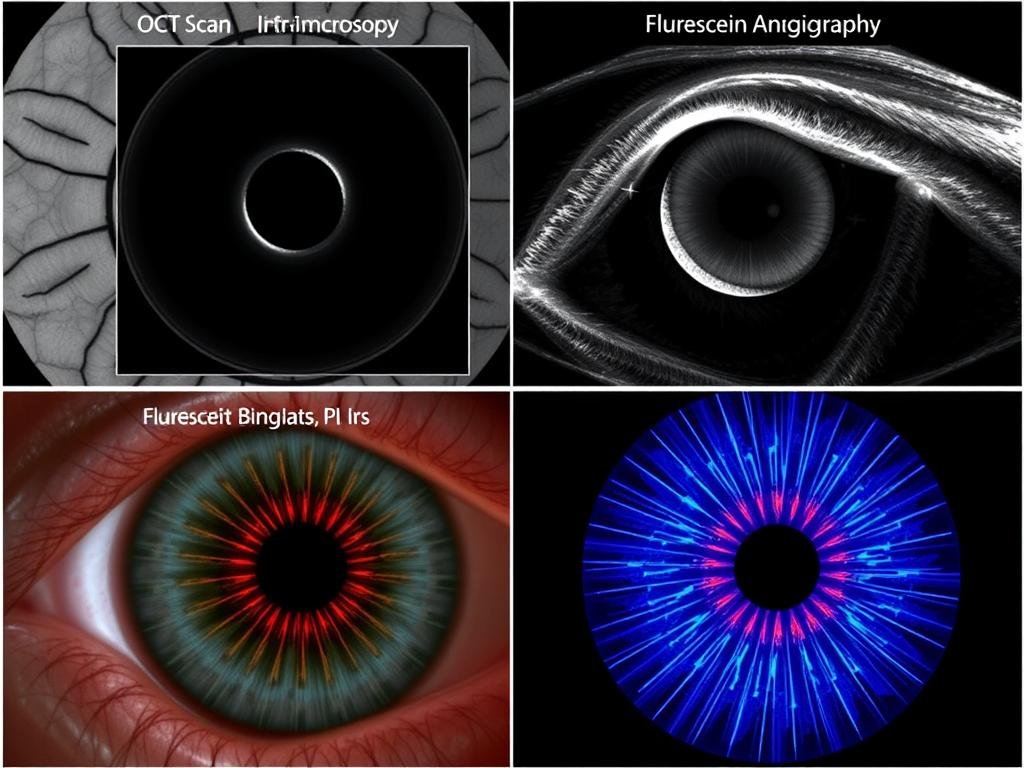
  • Optical Coherence Tomography (OCT)Creates cross-sectional images of the iris and surrounding structures
  • Ultrasound Biomicroscopy (UBM)Uses high-frequency sound waves to create detailed images of the iris and anterior chamber
  • Fluorescein AngiographyEvaluates blood flow in the iris by tracking a fluorescent dye

Advanced imaging of the iris medical term showing detailed structure

Advanced imaging techniques provide detailed views of iris structure

Frequently Asked Questions About the Iris

Can iris color change over time?

Most babies are born with blue or gray irises that may change color during their first year of life as melanin production increases. By age 3, iris color is usually stable. In adults, significant iris color changes are rare and may indicate an underlying condition requiring medical attention.

Are iris scanners harmful to the eyes?

Iris scanners used for biometric identification use low-level infrared light that is not harmful to the eyes. These devices capture the unique pattern of your iris without causing damage or discomfort. There is no scientific evidence suggesting that iris scanning technology poses any risk to eye health.

Why do pupils dilate during an eye exam?

Eye care professionals often use dilating drops to temporarily paralyze the iris muscles, keeping the pupil wide open. This dilation allows them to thoroughly examine the retina and other internal eye structures that would otherwise be difficult to see. The effect typically wears off within 4-6 hours.

Can iris color affect vision or light sensitivity?

Yes, people with lighter colored irises (blue, green) typically have less melanin pigment, which can make them more sensitive to bright light. Those with darker irises (brown) generally have more natural protection against glare and UV light. However, everyone should wear UV-protective sunglasses outdoors regardless of eye color.

What causes the pupil to appear red in photographs?

Ten “red-eyeeffect occurs when camera flash reflects off the blood-rich retina at the back of the eye. This happens when the pupil is dilated in low light and the flash is too quick for the iris to constrict. Many cameras have ared-eye reductionfeature that flashes twice—once to constrict the pupil and again to take the picture.

Glossary of Iris-Related Medical Terms

Anatomical Terms

  • UveaThe middle layer of the eye that includes the iris, ciliary body, and choroid
  • StromaThe fibrous tissue that forms the main body of the iris
  • Pupillary marginThe inner edge of the iris that forms the boundary of the pupil
  • CollaretteThe thickened ridge in the iris that marks the boundary between the pupillary and ciliary zones

Functional Terms

  • MiosisConstriction or narrowing of the pupil
  • MydriasisDilation or widening of the pupil
  • PhotophobiaAbnormal sensitivity to light, often due to iris inflammation
  • AnisocoriaUnequal pupil sizes, which may indicate an iris or neurological issue

Conditions

  • AniridiaCongenital absence of the iris, either partial or complete
  • IridocyclitisInflammation of both the iris and ciliary body
  • Rubeosis iridisAbnormal blood vessel growth on the iris surface
  • IridoschisisSplitting of the iris stroma, usually age-related

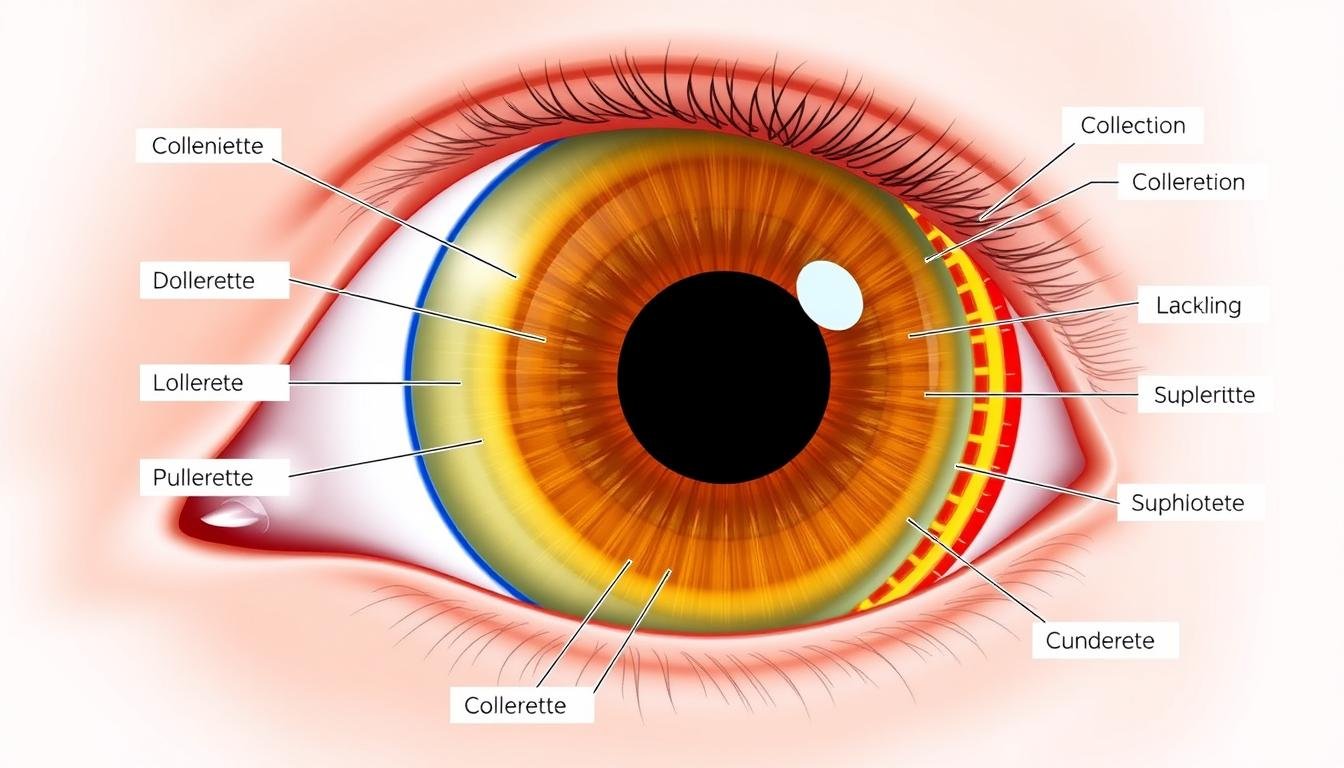
Medical diagram illustrating key iris medical term structures with labels

Key anatomical structures of the iris

References and Further Reading

Person reading about iris medical term on digital device with eye care professional

Consulting with eye care professionals is essential for maintaining iris health

Protecting Your Eye Health

Ten iris is a remarkable structure that not only gives your eyes their distinctive color but also plays a crucial role in vision by regulating light entry. Understanding the iris medical term and its functions can help you recognize potential issues and seek appropriate care when needed.

Regular eye examinations are essential for maintaining eye health and detecting conditions that may affect the iris before they cause vision problems. If you experience any changes in vision, eye pain, light sensitivity, or notice changes in your iris appearance, consult with an eye care specialist promptly.

Take Control of Your Eye Health Today

Consult an ophthalmologist if you experience light sensitivity or vision changes that could indicate iris-related conditions.

Schedule Your Eye Examination