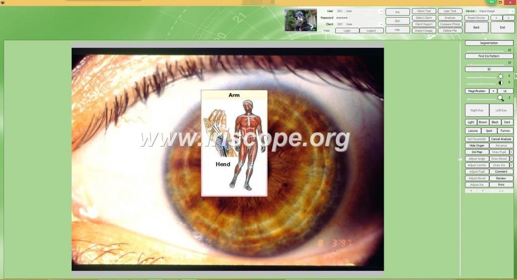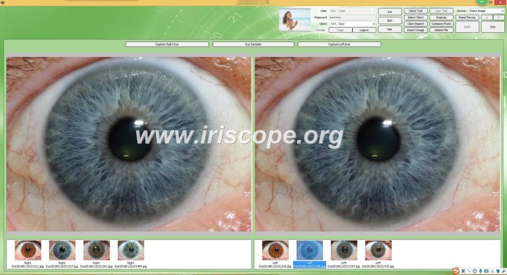

 iridology camera software how to use it?
1 Instalați software -ul.
2 Conexiune Orange USB Cheie Weith PC -ul dvs.
3 Deschideți desktop “CadiCV Advance Analysis System versiunea în engleză”
1) Utilizați selectați “utilizator”, Parolă: 111111 și faceți clic pe: “log in”
2) Faceți clic “instrument client”, introduceți informațiile despre clienți. Dacă ok, faceți clic “adăuga”, și clieck”aproape”
3) Faceți clic “capta ochiul drept”.–clic “capta”,
4) Repetarea ochiului stâng Ultimul pas.
5) Selectați imaginea ochiului (imaginea ochiului din dreapta / imaginea ochiului stâng)
6) Faceți clic “analiză”
7) Faceți clic “setați parametrul” buton.
8) Faceți clic “analiza irisului” buton.
9) Analiză
Observarea simptomelor și culoarea irisului la alegere în software -ul deasupra butonului corespunzător pe simptome și noapte.
De exemplu: aveți fisură pe imaginea irisului, iar culoarea este ușoară.
Selectați butonul „fisură” și butonul „lumină”, mutați cursorul pe fisurile de pe iris,
Faceți clic cu mouse -ul. Raportat imediat prin analiză.
Tu și adăugați recomandarea sau produsul pentru client.
adăuga ——? Analiză——–? stânga (Analiza ochiului stâng)
10 Salvați
11 Raport - Selectați numele raportului —–? Raport HTML sau raport Privew Selectați numele rapoartelor (numele datei)
12 tipăriri
13 Ștergeți clientul
14 Editați clientul


Iridologist Job Duties
Those considering careers in iridology should note that iridology is not recognized by the mainstream medical community in the U.S. A study published in the Journal of the American Medical Association showed evidence that iridology has no therapeutic value, and most U.S. insurance companies do not cover iridology.According to the International Iridology Practitioners Association (IIPA), iridologists conduct close examinations of the human iris, the colored portion of the eye, using tools such as flashlights, magnifying glasses and cameras . After taking close-up images of the iris, iridologists examine factors such as eye color, spots and noticeable rings. They also look at the white portion of the eyes, known as the sclera. Iridologists then compare images of the patient’s eyes to various charts that indicate correlations between markers on the iris and sclera with potential health conditions.During consultations with patients, iridologists explain any abnormalities found on the patient’s iris or sclera. For example, certain iris colors have been linked to digestive problems or allergies. Iridologists may instruct patients on homeopathic ways of dealing with health problems, such as taking supplements, making dietary changes or increasing physical activity.
How to take your iridology images by irideology camera software?
Use your thumb to pull down your bottom eyelid, while at the same time use your index finger to lift-up your top eyelid until the entire eye is visible (the top and bottom part of your eye must be visible); if that doesn’t work, then open your eye wide enough to where the photographer can see the top and bottom part of your eye. Then Look Straight into the camera’s eye, and shine a flashlight, or pin light directly at each corner of your eye (one eye at a time). This will illuminate your iris fibers so that the camera can pick-up the iris fibers. All is needed is a photo of your iris (eye) and not your entire face .


