The human iris contains over 28,000 nerve fibers connected to every organ and tissue in the body through the brain and nervous system. Iridology cameras are specialized imaging devices designed to capture high-resolution photographs of the iris, enabling health practitioners to analyze these intricate patterns for potential health insights. These advanced optical instruments have revolutionized the practice of علم القزحية by providing unprecedented detail and accuracy in iris examination.
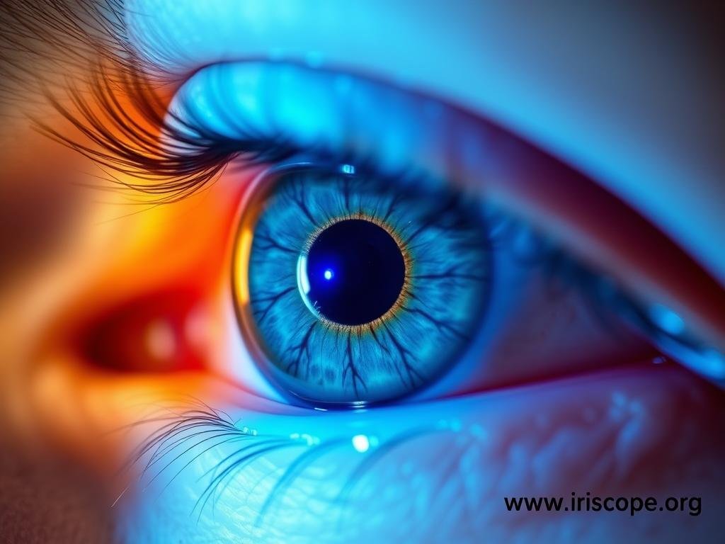
Advanced iridology camera with specialized lighting system for optimal iris imaging
Iridology cameras combine precision optics, specialized lighting, and digital imaging technology to capture the intricate details of the iris. Unlike conventional cameras, these specialized devices must overcome several unique challenges to produce clinically useful images. The iris is a moving, three-dimensional structure behind the curved cornea, requiring specific optical solutions to capture clear, undistorted images.

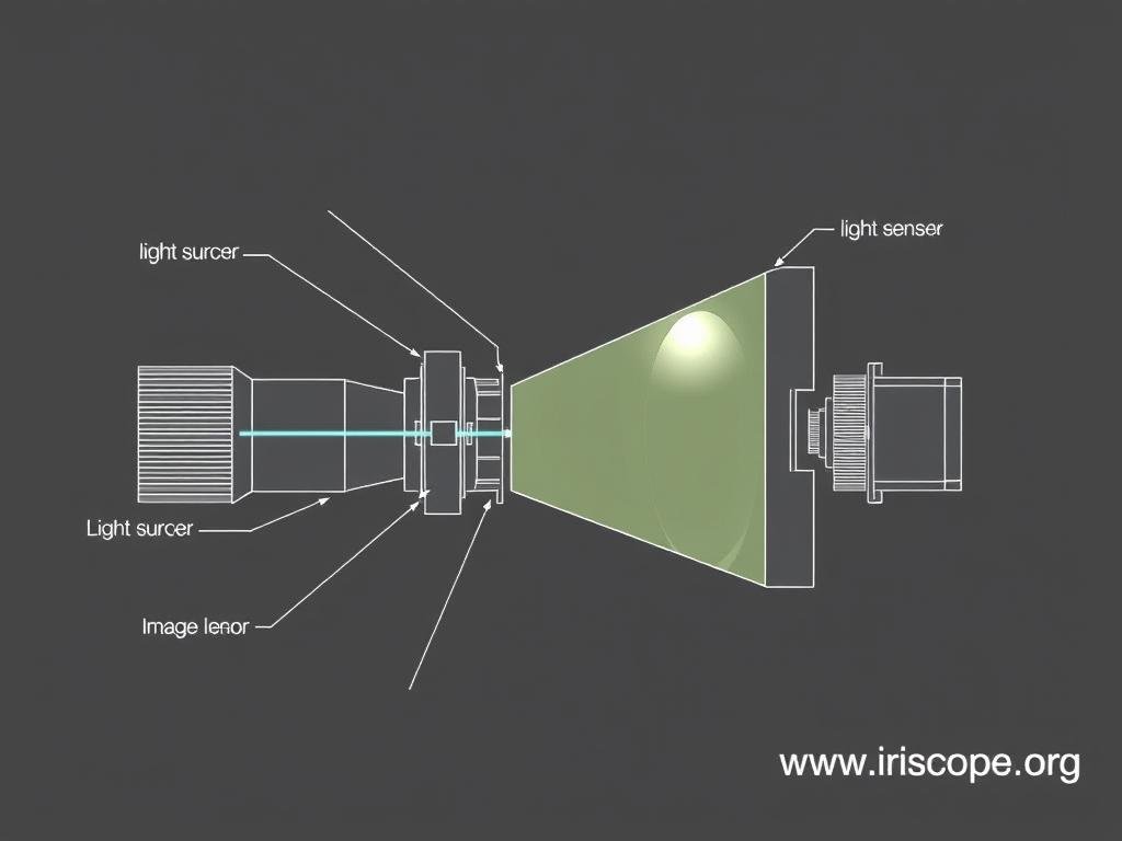
Optical pathway diagram of modern iridology camera systems
Key Components of Modern Iridology Cameras
Every professional iridology camera system integrates several critical components that work together to produce diagnostic-quality iris images:
- High-resolution image sensors (typically 12-24 megapixels)
- Specialized macro lenses with optimal focal length
- Adjustable, diffused lighting systems
- Precise focus control mechanisms
- Image processing software for enhancement and analysis
- Ergonomic housing for stable positioning

Precision optics in professional-grade iridology camera
Lighting Technology in Iridology Cameras
Lighting is perhaps the most critical element in iridology camera design. The ideal system provides even, diffused illumination that reveals iris details without causing pupil constriction or patient discomfort. Modern cameras employ various lighting configurations:
Lighting Type |
وصف |
Advantages |
Best For |
Twin Side Lighting |
Dual light sources positioned at 45° angles |
Even illumination, reduced reflections |
General iris examination |
Single Side Lighting |
One directional light source |
Enhanced topographical details |
Examining iris relief and depth |
Diffused LED Array |
Multiple small LEDs in circular arrangement |
Minimal shadows, consistent illumination |
Dark brown irises |
Cold Light Fiber Optics |
Remote light source delivered via fiber optics |
No heat transfer, patient comfort |
Extended examination sessions |
Digital vs. Analog Iridology Cameras
The evolution from analog to digital iridology cameras represents a significant technological advancement in the field. Understanding the differences helps practitioners select the most appropriate system for their needs.
أنظمة علم القزحية الرقمية
- Immediate image preview and capture
- No film or development costs
- Direct computer integration for analysis
- Higher resolution (12-24 megapixels)
- Adjustable settings for different iris types
- Image enhancement capabilities
- Easy storage and retrieval of patient records
Analog Iridology Systems
- Requires film and development process
- No immediate image review
- Limited number of exposures per session
- Manual record-keeping system
- Consistent image quality (no digital artifacts)
- No battery dependency
- Often more durable in harsh conditions

Evolution of technology: Modern digital iridology camera (left) compared to traditional analog system (right)
USB and Wireless Iridology Cameras
The latest generation of iridology cameras offers enhanced connectivity options that streamline the examination process:
USB Iridology Systems
- Direct computer connection for immediate image transfer
- Powered through USB connection (no separate power supply)
- Compatible with most analysis software
- Typically more affordable than wireless options
- Simple plug-and-play operation
Wireless Iridology Systems
- Freedom of movement during examination
- Built-in memory card for standalone operation
- Rechargeable battery power
- Wi-Fi or Bluetooth connectivity
- Ideal for mobile practitioners

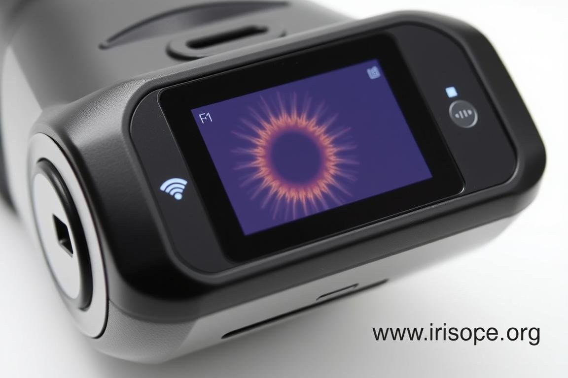
Portable wireless iridology camera with built-in display and memory storage
Technical Specifications of Professional Iridology Cameras
When evaluating iridology cameras for professional use, several key technical specifications determine image quality and clinical utility:
4.8
Critical Factors in Camera Selection
Optical Magnification
4.7
Resolution and Image Quality in Iridology Cameras
The resolution of iridology cameras directly impacts the level of detail visible in iris images. Professional systems typically offer:
Resolution Category |
Megapixels |
Typical Image Size |
Clinical Application |
Standard |
5-8 MP |
2560×1920 px |
Basic screening, general patterns |
Professional |
12-18 MP |
4032×3024 px |
Detailed analysis, clinical practice |
Research Grade |
24-36 MP |
6000×4000 px |
Advanced research, minute details |

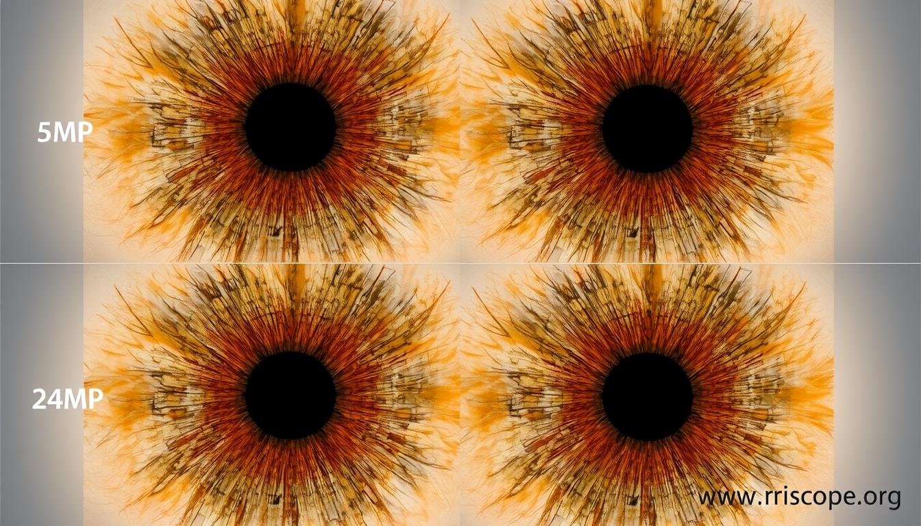
Comparison of iris images captured with iridology cameras at different resolutions: 5MP (left), 12MP (center), 24MP (right)
Clinical Applications of Iridology Cameras
Professional iridology cameras serve multiple purposes in clinical practice, extending beyond traditional iridology to various health assessment applications:
تقييم الصحة الشاملة
Practitioners use high-resolution iris images to identify constitutional strengths and weaknesses, supporting personalized health recommendations.
Patient Education
Displaying detailed iris images helps patients visualize and understand their health patterns, improving compliance with recommended protocols.
Progress Monitoring
Sequential iris imaging allows practitioners to document changes over time, providing visual evidence of health improvements or challenges.
Specialized Applications of Iridology Cameras
Beyond standard iridology, these advanced iridology cameras support several specialized examination techniques:
Sclerology
Examining the blood vessels and markings in the sclera (white of the eye) provides additional health insights. Modern iridology cameras with adjustable lighting are ideal for capturing both iris and sclera details in the same session.

Sclera examination using specialized lighting settings on a professional iridology camera
“The advancement in iridology camera technology has transformed our clinical practice. We can now detect subtle changes in iris structure that were previously invisible, allowing for more precise and personalized health recommendations.”
– Dr. Maria Chen, Naturopathic Physician
Maintenance and Care of Iridology Cameras
Professional iridology cameras represent a significant investment and require proper maintenance to ensure optimal performance and longevity:
Essential Maintenance Practices
- Clean optical surfaces using only approved lens cleaning solutions and microfiber cloths
- Store the camera in a dust-free, temperature-controlled environment
- Regularly check and clean lighting elements to maintain consistent illumination
- Update camera firmware and associated software when available
- Calibrate color and white balance settings periodically
- Inspect all cables and connections for wear or damage
- Schedule professional servicing annually for optimal performance

Proper cleaning technique for iridology camera optics using appropriate tools
Important: Never use alcohol-based cleaners directly on camera lenses or illumination systems as they can damage coatings and seals. Always use products specifically designed for optical equipment.
اختيار كاميرا علم القزحية المناسبة لعيادتك
Selecting the appropriate iridology camera depends on several factors including your clinical needs, budget, and technical requirements:
What resolution is necessary for professional iridology practice?
For professional clinical practice, we recommend a minimum of 12 megapixels. This resolution provides sufficient detail to observe fine iris structures while keeping file sizes manageable. Research-oriented practices may benefit from higher resolutions (24+ MP) for examining minute details.
Are portable or stationary iridology camera systems better?
This depends on your practice model. Stationary systems typically offer higher stability and consistent results but require dedicated space. Portable systems provide flexibility for multiple office locations or home visits but may sacrifice some image stability. Many practitioners choose a portable system with an optional mounting stand for the best of both worlds.
How important is specialized software with an iridology camera?
Integrated software significantly enhances the clinical utility of iridology cameras. Look for systems that offer iris mapping, comparison tools, client records management, and reporting features. The best software allows for iris chart overlays, measurement tools, and image enhancement capabilities.

Range of iridology camera options from entry-level to advanced professional systems
Advancing Your Practice with Professional Iridology Cameras
Iridology cameras have evolved from simple magnifying tools to sophisticated digital imaging systems that enhance clinical practice and patient education. By understanding the technical aspects, maintenance requirements, and clinical applications of these specialized devices, practitioners can make informed decisions when investing in this essential equipment.
Whether you’re establishing a new practice or upgrading existing equipment, selecting the right iridology camera system is crucial for providing accurate assessments and maintaining professional standards. The technology continues to advance, offering increasingly detailed insights into the fascinating world of iris analysis.
Expert Iridology Camera Solutions
We provide professional-grade iridology cameras with comprehensive technical support and training. Contact our specialists to discuss your specific requirements and find the perfect system for your practice.
Contact on WhatsApp
Email Us