المصطلح الطبي القزحية: القزحية هي البنية الدائرية الملونة في عينك والتي تحيط بالبؤبؤ. باعتبارها عنصرًا حاسمًا في تشريح العين، تتحكم القزحية في مقدار الضوء الذي يدخل عينك عن طريق ضبط حجم حدقة العين. هذا الهيكل الرائع لا يمنح عينيك لونها المميز فحسب، بل يلعب أيضًا دورًا حيويًا في الرؤية من خلال تنظيم تعرض شبكية العين للضوء. في هذا الدليل الشامل، سنستكشف بنية القزحية ووظائفها والظروف المرتبطة بها وأهميتها في فحوصات العين.
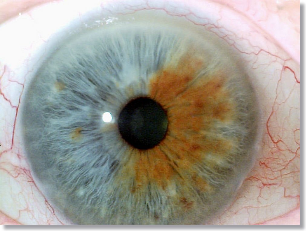
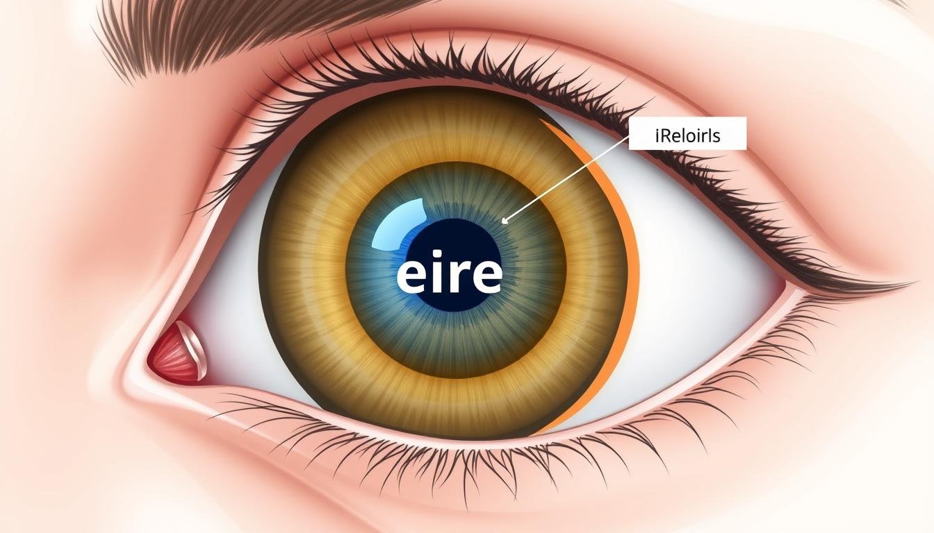
مقطع عرضي تشريحي يوضح موقع القزحية بين القرنية والعدسة
ما هي القزحية؟ فهم بنية العين الأساسية هذه
المصطلح الطبي القزحية: القزحية هي البنية الدائرية الرفيعة في العين المسؤولة عن التحكم في قطر وحجم بؤبؤ العين. تقع القزحية بين القرنية والعدسة، وتفصل الحجرة الأمامية عن الغرفة الخلفية للعين. الكلمة “قزحية” مشتق من الكلمة اليونانية ل “قوس قزح،” مما يعكس الألوان المتنوعة التي يمكن أن يعرضها هذا الهيكل عند البشر.
من الناحية التشريحية، القزحية هي جزء من القناة العنبية، والتي تشمل أيضًا الجسم المشيمي والجسم الهدبي. ويمتد عبر الجزء الأمامي من العين ويتصل بالجسم الهدبي عند حافته الخارجية (المعروف باسم جذر القزحية). وتشكل الفتحة المركزية للقزحية حدقة العين، التي تظهر باللون الأسود لأن الضوء الذي يمر عبرها تمتصه الأنسجة الموجودة داخل العين.
تعمل القزحية بمثابة نظام فتحة العين الطبيعية، على غرار الحجاب الحاجز في الكاميرا. من خلال التحكم في مقدار الضوء الذي يصل إلى شبكية العين، فإنه يساعد على تحسين الرؤية في ظروف الإضاءة المختلفة ويحمي أنسجة الشبكية الحساسة من الأضرار المحتملة الناجمة عن التعرض المفرط للضوء.
تشعر بالقلق إزاء صحة عينيك؟
تعد فحوصات العين المنتظمة ضرورية للحفاظ على الرؤية المثالية واكتشاف المشكلات المحتملة مبكرًا.
حدد موعدًا لفحص العين
بنية القزحية: التشريح المعقد للجزء الملون من عينك
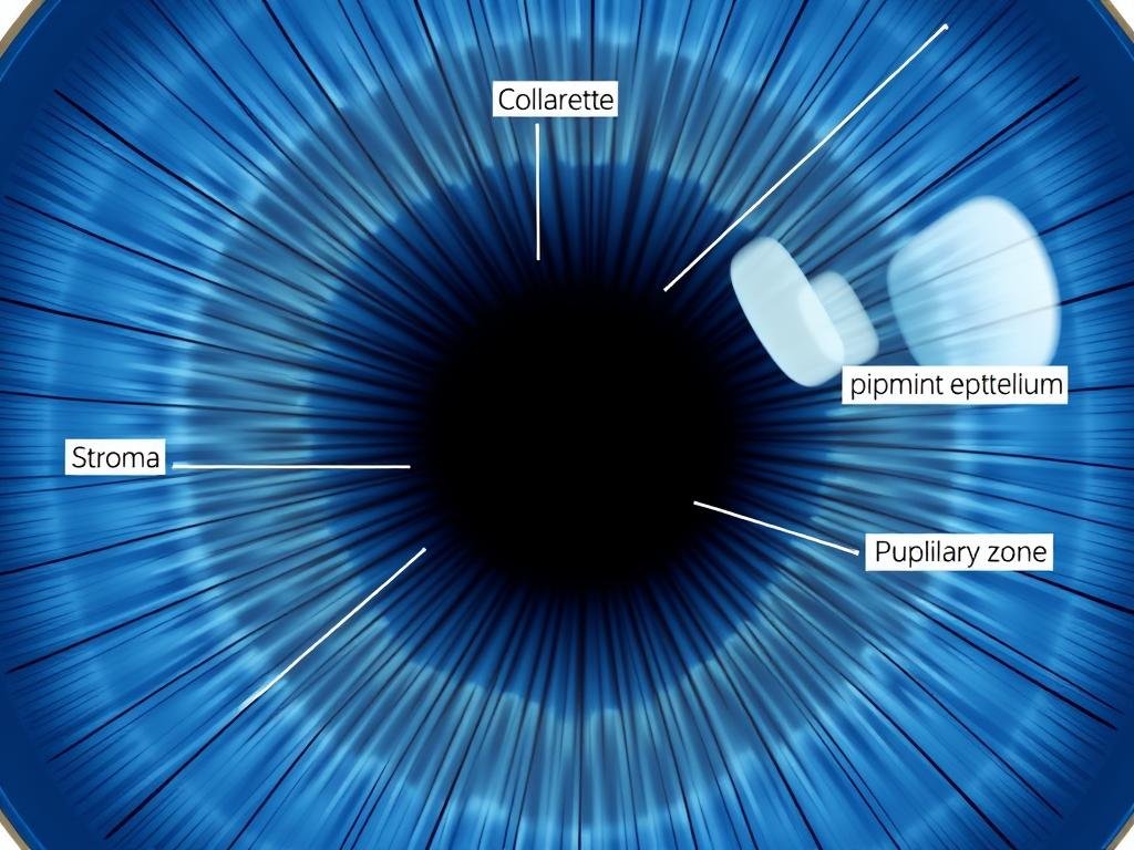
عرض تفصيلي لهياكل القزحية بما في ذلك الياقة والسدى ومنطقة الحدقة
تمتلك القزحية بنية معقدة تتكون من عدة طبقات ومناطق متميزة. يساعد فهم هذه المكونات في تفسير كيفية عمل هذا النسيج الرائع ويؤدي إلى ظهور مجموعة واسعة من ألوان العين التي نلاحظها عند البشر.
المكونات الهيكلية الرئيسية للقزحية
المناطق والأقاليم
تنقسم القزحية إلى منطقتين أساسيتين:
- منطقة الحدقة – المنطقة الداخلية التي تشكل حدود التلميذ
- المنطقة الهدبية – المنطقة الخارجية الممتدة إلى جذر القزحية عند الجسم الهدبي
ال طوق هي الحافة السميكة التي تفصل بين هاتين المنطقتين. وهو يمثل بقايا الغشاء الحدقي الجنيني ويظهر كدائرة غير منتظمة قليلاً بالقرب من هامش الحدقة.
طبقات الأنسجة
من الأمام إلى الخلف، تتكون القزحية من:
- طبقة الحدود الأمامية – السطح الأمامي يحتوي على الخلايا الصباغية والخلايا الليفية
- سدى – طبقة من النسيج الضام فضفاضة مع الأوعية الدموية والأعصاب والخلايا الصباغية
- العضلات العاصرة والموسعة – السيطرة على حجم التلميذ
- ظهارة الصباغ – طبقتان من الخلايا المصطبغة بشدة على السطح الخلفي
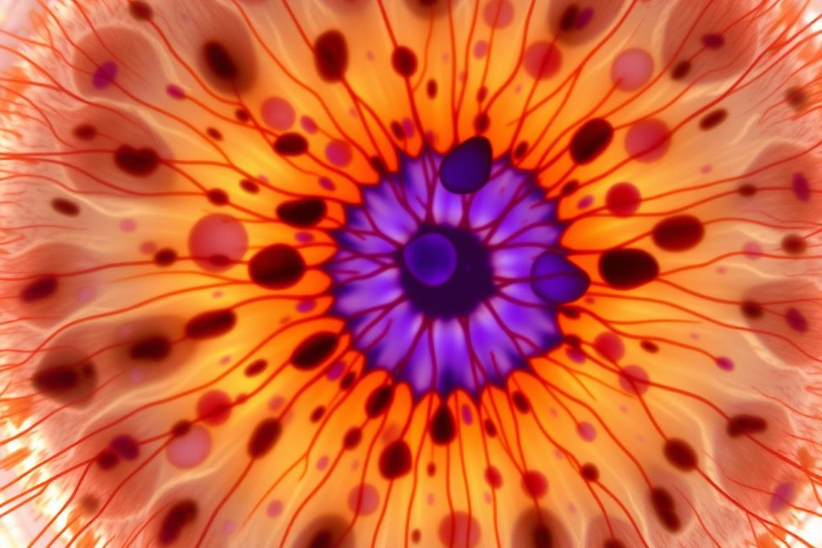
منظر مجهري لأنسجة القزحية تظهر الخلايا الصباغية وتوزيع الصباغ
السدى: مركز لون القزحية
السدى عبارة عن طبقة ليفية إسفنجية تحتوي على الخلايا الصباغية - وهي خلايا متخصصة تنتج صبغة الميلانين. تحدد كمية وتوزيع الميلانين في السدى لون عينيك إلى حد كبير. تحتوي العيون البنية على وفرة من الميلانين في جميع أنحاء السدى، بينما تحتوي العيون الزرقاء على الحد الأدنى من الميلانين اللحمي، مما يتسبب في تبعثر الضوء وانعكاسه (على غرار كيف تبدو السماء زرقاء).
ظهارة الصباغ: خلفية القزحية
المصطلح الطبي القزحية: تشكل الظهارة الصبغية الخلفية طبقة كثيفة غير شفافة تمنع دخول الضوء إلى العين إلا من خلال حدقة العين. تحتوي هذه الطبقة على صبغة الميلانين الثقيلة بغض النظر عن لون العين، ولهذا السبب حتى الأشخاص ذوي العيون الفاتحة لا يعانون من تسرب الضوء عبر أنسجة القزحية.
| ميزة |
قزحية |
القرنية |
شبكية العين |
| الوظيفة الأساسية |
يتحكم في دخول الضوء عن طريق ضبط حجم حدقة العين |
يعكس الضوء الذي يدخل إلى العين |
تحويل الضوء إلى إشارات عصبية |
| موقع |
بين القرنية والعدسة |
السطح الأمامي للعين |
السطح الخلفي الداخلي للعين |
| نوع الأنسجة |
عضلي ومصطبغ |
شفاف، غير وعائي |
الأنسجة العصبية مع المستقبلات الضوئية |
| إمدادات الدم |
شديد الأوعية الدموية |
لا وعائي (لا توجد أوعية دموية) |
شبكة معقدة من أوعية الشبكية |
| الاضطرابات الشائعة |
التهاب القزحية، تغاير اللون، أنيريديا |
التهاب القرنية، تقرحات القرنية، القرنية المخروطية |
انفصال الشبكية، الضمور البقعي |
وظائف القزحية: أكثر من مجرد لون العين
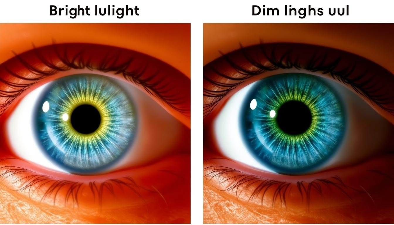
إظهار وظيفة القزحية: انقباض حدقة العين في الضوء الساطع (يسار) مقابل تمددها في الضوء الخافت (يمين)
في حين أن العديد من الناس يربطون القزحية في المقام الأول بلون العين، فإن هذا الهيكل الرائع يؤدي العديد من الوظائف الحيوية التي تعتبر ضرورية للرؤية وصحة العين.
تنظيم الضوء: الدور الأساسي للقزحية
أهم وظيفة للقزحية هي التحكم في كمية الضوء التي تدخل العين. ويحقق ذلك من خلال مجموعتين من العضلات:
- العضلة العاصرة الحدقة – عضلة دائرية تنقبض لتضييق حدقة العين في الظروف الساطعة
- موسع الحدقة – العضلات الشعاعية التي تعمل على توسيع حدقة العين في الظروف المظلمة
This dynamic adjustment helps optimize vision across varying light conditions. In bright environments, pupil constriction prevents the retina from being overwhelmed by excessive light. In dim settings, pupil dilation allows maximum light collection for better visibility.
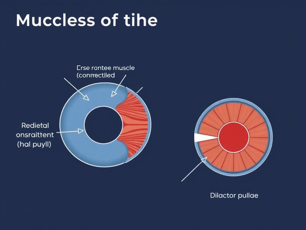
Illustration of iris muscles: sphincter pupillae (circular) and dilator pupillae (radial)
Additional Functions of the Iris
UV Protection
The melanin pigment in the iris helps absorb ultraviolet radiation, providing a protective function for the internal structures of the eye. People with lighter iris colors have less melanin and may be more susceptible to light-related eye damage.
Depth of Field Enhancement
When you focus on nearby objects, your pupils constrict in a process called the accommodative pupillary response. This increases your depth of field (the range of distances that appear in focus), improving visual clarity for close-up tasks.
Aesthetic and Identification Functions
Medical Term Iris: Beyond its physiological roles, the iris provides each person with a unique eye color and pattern. Iris patterns are so distinctive that they’re used in biometric identification systems. The diversity of iris colors—from brown, blue, and green to hazel, gray, and amber—results from variations in melanin concentration and distribution within the iris stroma.
الحالات والاضطرابات المتعلقة بالقزحية
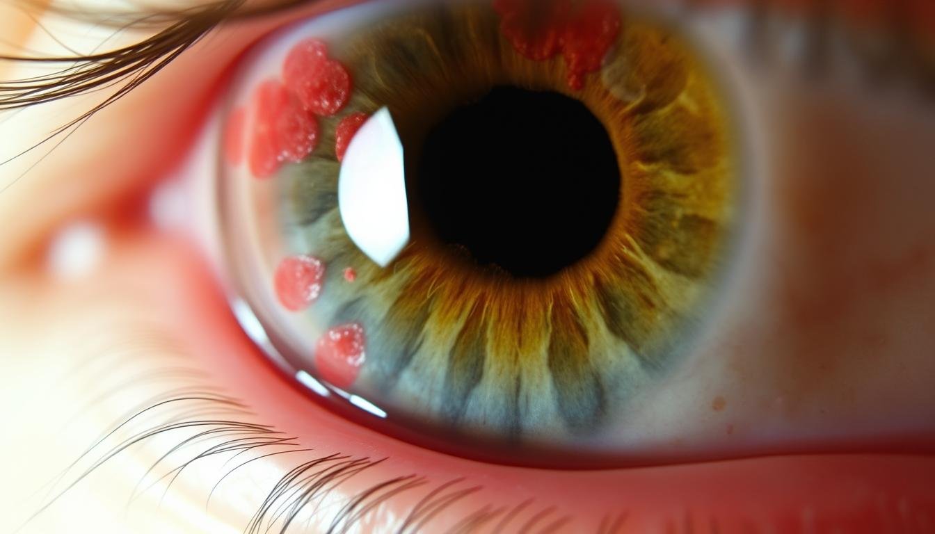
العرض السريري لالتهاب القزحية الحاد يظهر التهاب وحدقة غير منتظمة
يمكن أن تؤثر عدة حالات على القزحية، بدءًا من الاضطرابات الالتهابية وحتى التشوهات الخلقية. إن فهم هذه الحالات مهم للكشف المبكر والعلاج.
الحالات الالتهابية
التهاب القزحية (التهاب العنبية الأمامي)
Iritis is inflammation of the iris and anterior chamber of the eye. It can cause pain, light sensitivity, blurred vision, and a small, irregular pupil. Acute iritis often develops suddenly and may be associated with autoimmune disorders, infections, or trauma. According to a study published in the Journal of Ophthalmology, iritis affects approximately 12 per 100,000 people annually.
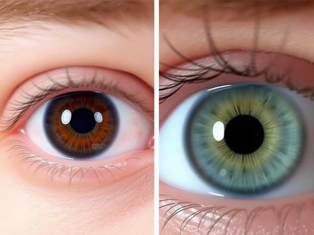
Complete heterochromia: different colored irises in the same individual
Pigmentation Disorders
تغاير اللون
Heterochromia is a condition where a person has different colored irises or different colors within the same iris. Complete heterochromia (heterochromia iridis) involves two differently colored eyes, while partial heterochromia (heterochromia iridium) involves different colors within the same iris. This condition can be congenital or acquired due to injury, inflammation, or certain medications.
المهق العيني
Ocular albinism affects the pigmentation of the eyes without necessarily affecting skin and hair color. People with this condition typically have very light-colored irises that appear translucent due to reduced melanin. This can cause increased light sensitivity and reduced visual acuity. Research in the American Journal of Ophthalmology indicates that ocular albinism affects approximately 1 in 60,000 males.
Structural Abnormalities
أنيريديا
Aniridia is a rare congenital condition characterized by the partial or complete absence of the iris. People with aniridia typically have poor vision, light sensitivity, and are at increased risk for other eye problems like glaucoma and cataracts. The condition affects approximately 1 in 50,000 to 100,000 people worldwide.
كولوبوما
An iris coloboma appears as a keyhole-shaped pupil due to incomplete formation of the iris during embryonic development. This gap in the iris structure can cause increased light sensitivity and blurred vision. Colobomas may occur as isolated findings or as part of genetic syndromes.
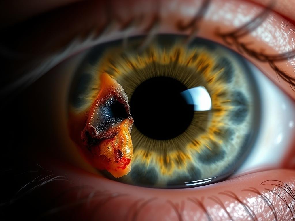
Traumatic injury to the iris showing structural damage and irregular pupil
Traumatic Injuries
ال قزحية can be damaged by blunt or penetrating eye injuries. Common traumatic iris conditions include:
- Iridodialysis – Separation of the iris from its attachment at the ciliary body
- Traumatic mydriasis – Permanent pupil dilation due to sphincter muscle damage
- Iris tears – Ruptures in the iris tissue that may affect pupillary function
According to a study in the British Journal of Ophthalmology, approximately 15% of all eye injuries involve damage to the iris structure.
Experiencing Eye Problems?
If you’re experiencing changes in vision, eye pain, or unusual iris appearance, consult with an eye specialist promptly.
Learn More About Iris Conditions
Diagnostic Importance of the Iris
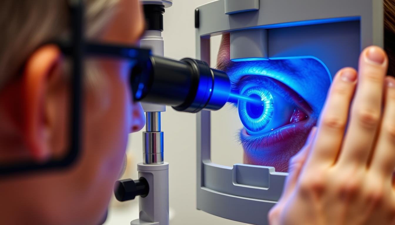
Ophthalmologist examining the iris using a slit lamp biomicroscope
The iris plays a significant role in various diagnostic assessments, providing valuable information about ocular and systemic health.
Iris Examination in Routine Eye Care
During a comprehensive eye examination, eye care professionals carefully assess the iris for:
- Color and pigmentation patterns
- Structural integrity and symmetry
- Pupillary responses to light and accommodation
- Signs of inflammation or abnormal blood vessels
- Presence of nodules, cysts, or other growths
These observations can reveal early signs of ocular conditions or systemic diseases that manifest in the iris. For example, certain inflammatory patterns may suggest autoimmune disorders, while specific iris changes might indicate diabetes or hypertension.
Neurological Assessment
The pupillary light reflex—controlled by the iris muscles—is a critical component of neurological examinations. Abnormal pupillary responses can indicate various neurological conditions, including:
- Brain injuries or increased intracranial pressure
- Oculomotor nerve (CN III) dysfunction
- Horner’s syndrome
- Effects of certain medications or substances
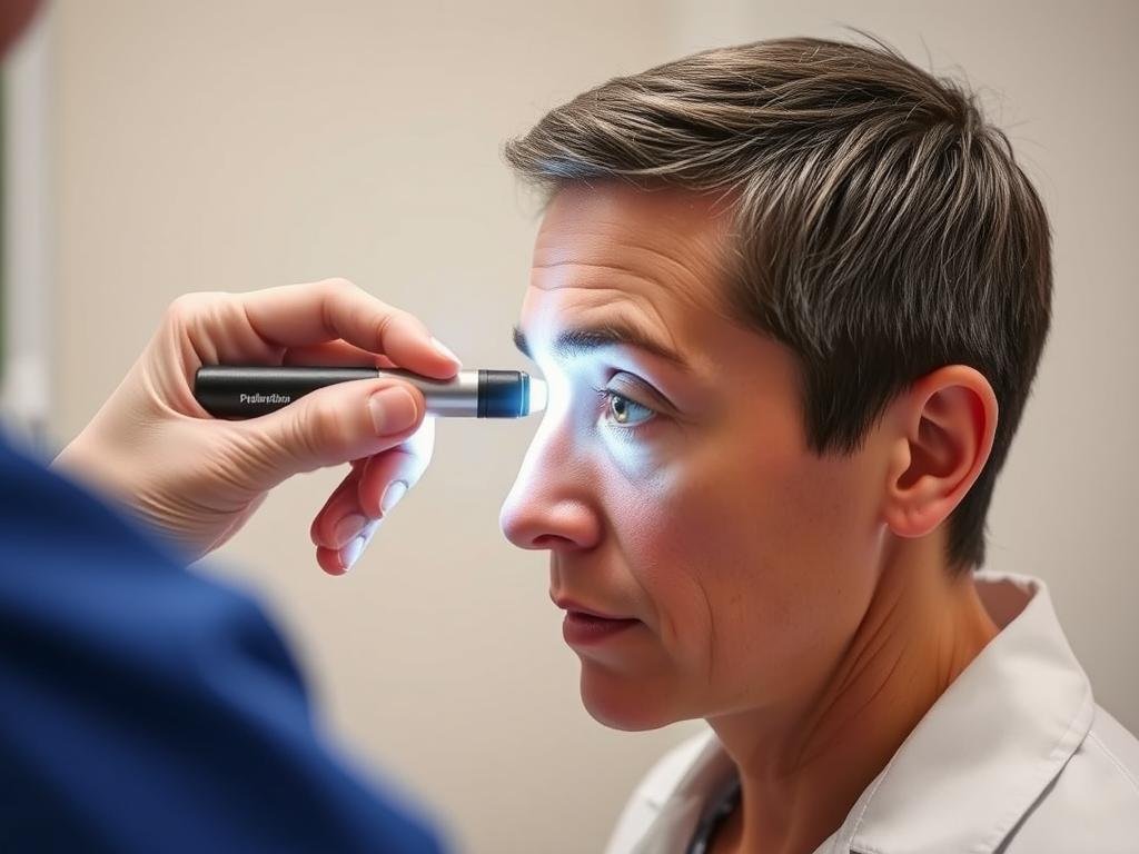
Pupillary light reflex testing – an important neurological assessment of iris function
According to research published in the Journal of Neurology, pupillary assessment has a sensitivity of over 90% for detecting certain types of brain injuries when performed correctly.
Maintain Your Eye Health
Regular comprehensive eye examinations are essential for early detection of eye conditions and maintaining optimal vision.
حدد موعدًا لفحص عينيك اليوم
Frequently Asked Questions About the Iris
Can iris color change throughout life?
While most people’s iris color stabilizes by age 3, some gradual changes can occur throughout life. Babies, particularly those of Caucasian descent, are often born with blue or gray irises that may darken during early childhood as melanin production increases. In adults, certain medications (like prostaglandin analogs used for glaucoma) can gradually darken iris color. Additionally, some eye conditions or injuries may alter iris appearance. However, dramatic spontaneous changes in adult iris color are rare and should be evaluated by an eye care professional.
What causes heterochromia (different colored irises)?
Medical Term Iris: Heterochromia can be congenital (present from birth) or acquired later in life. Congenital heterochromia is typically caused by genetic mutations affecting melanin production or distribution in the iris. It may occur as an isolated finding or as part of syndromes like Waardenburg syndrome. Acquired heterochromia can result from eye injuries, certain medications, inflammation (like chronic iritis), or conditions such as Horner’s syndrome. While often harmless, new-onset heterochromia should be evaluated by an eye specialist to rule out underlying conditions.
How does iritis (iris inflammation) affect vision?
Iritis can significantly impact vision through several mechanisms. The inflammation causes light sensitivity (photophobia) and pain, making it difficult to function in normal lighting conditions. Inflammatory cells in the anterior chamber can cloud vision, while inflammatory debris can settle on the lens, causing blurring. The inflamed iris may also stick to the lens (posterior synechiae), affecting pupil function and potentially leading to complications like glaucoma or cataracts. Prompt treatment with anti-inflammatory medications is essential to prevent permanent vision damage.
Can iris damage be repaired?
Treatment options for iris damage depend on the type and extent of injury. Minor iris tears may heal without intervention, while more significant damage might require surgical repair. Iridoplasty can reshape the iris, while iris prostheses or implants can address larger defects. However, not all iris damage can be fully restored to pre-injury appearance or function. Modern surgical techniques have improved outcomes for traumatic iris injuries, but prevention through protective eyewear remains the best approach. Any iris injury should receive prompt medical attention to minimize complications.
What does it mean if my pupil doesn’t respond normally to light?
Medical Term Iris: Abnormal pupillary responses can indicate various conditions affecting the iris muscles, nerves, or brain. A sluggish or absent pupillary light reflex might suggest oculomotor nerve damage, while unequal pupil sizes (anisocoria) could indicate Horner’s syndrome, Adie’s tonic pupil, or medication effects. In emergency settings, abnormal pupillary responses may signal increased intracranial pressure or brain injury. Certain medications and eye drops can also affect pupil reactivity. Any new or unexplained change in pupillary response warrants prompt medical evaluation, as it could represent anything from a benign condition to a medical emergency.
Understanding Your Iris for Better Eye Health
The iris is much more than just the colored part of your eye—it’s a complex structure that plays a crucial role in vision and can provide important insights into your overall health. From regulating light entry to creating your unique eye color, the iris performs several vital functions that contribute to clear vision and eye protection.
By understanding normal iris anatomy and function, you can better recognize potential problems and seek appropriate care when needed. Regular comprehensive eye examinations are essential for monitoring iris health and detecting conditions early, when they’re most treatable.
If you notice any changes in your iris appearance, pupil function, or vision, consult with an eye care professional promptly. With proper care and attention, you can help maintain the health of this remarkable structure and protect your vision for years to come.


