Medical Term Iris : The iris is the colored circular structure in your eye that surrounds the pupil. As a critical component of ocular anatomy, the iris controls how much light enters your eye by adjusting the size of your pupil. This remarkable structure not only gives your eyes their distinctive color but also plays a vital role in vision by regulating light exposure to the retina. In this comprehensive guide, we’ll explore the iris’s structure, functions, associated conditions, and its importance in eye examinations.

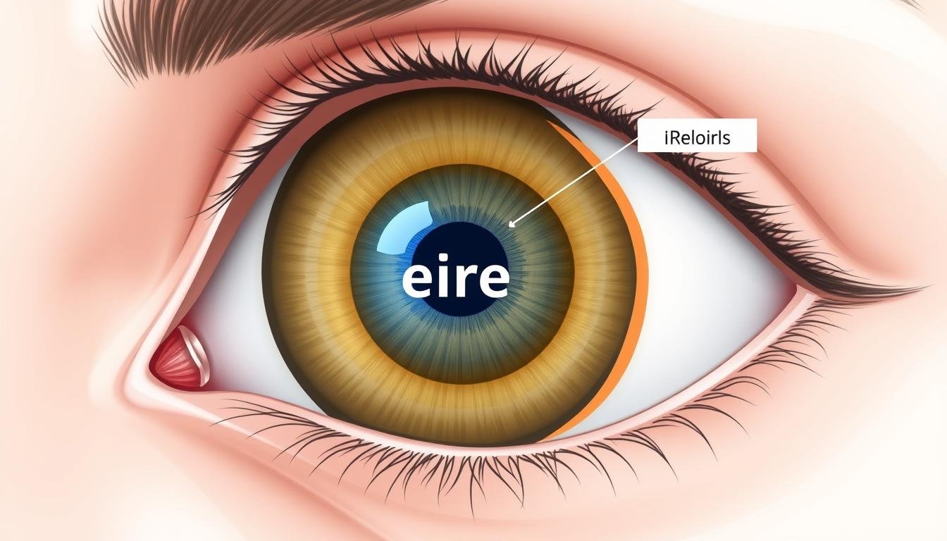
Anatomical cross-section showing the iris’s position between the cornea and lens
What Is the Iris? Understanding This Essential Eye Structure
Medical Term Iris: The iris is the thin, circular structure in the eye responsible for controlling the diameter and size of the pupil. Located between the cornea and the lens, the iris divides the anterior chamber from the posterior chamber of the eye. The word “iris” derives from the Greek word for “rainbow,” reflecting the varied colors this structure can display in humans.
Anatomically, the iris is part of the uveal tract, which also includes the choroid and ciliary body. It extends across the anterior portion of the eye and connects to the ciliary body at its outer edge (known as the iris root). The central opening of the iris forms the pupil, which appears black because light passing through it is absorbed by the tissues inside the eye.
The iris serves as the eye’s natural aperture system, similar to the diaphragm in a camera. By controlling how much light reaches the retina, it helps optimize vision in varying light conditions and protects the sensitive retinal tissue from potential damage caused by excessive light exposure.
Préoccupé par votre santé oculaire?
Regular eye examinations are essential for maintaining optimal vision and detecting potential issues early.
Schedule an Eye Examination
Iris Structure: The Complex Anatomy of Your Eye’s Colored Part

Detailed view of iris structures including the collarette, stroma, and pupillary zone
The iris has a complex structure composed of several distinct layers and regions. Understanding these components helps explain how this remarkable tissue functions and gives rise to the wide variety of eye colors we observe in humans.
Key Structural Components of the Iris
Zones et régions
The iris is divided into two primary zones:
- Pupillary zone – The inner region that forms the boundary of the pupil
- Ciliary zone – The outer region extending to the iris root at the ciliary body
Le collarette is the thickened ridge that separates these two zones. It represents a vestige of the embryonic pupillary membrane and appears as a slightly irregular circle near the pupillary margin.
Tissue Layers
From front to back, the iris consists of:
- Anterior border layer – The front surface containing melanocytes and fibroblasts
- Stroma – A loose connective tissue layer with blood vessels, nerves, and melanocytes
- Sphincter and dilator muscles – Control pupil size
- Pigment epithelium – Two layers of heavily pigmented cells at the back surface

Microscopic view of iris tissue showing melanocytes and pigment distribution
The Stroma: Center of Iris Color
The stroma is a spongy, fibrovascular layer that contains melanocytes—specialized cells that produce the pigment melanin. The amount and distribution of melanin in the stroma largely determines your eye color. Brown eyes have abundant melanin throughout the stroma, while blue eyes have minimal stromal melanin, causing light to scatter and reflect (similar to how the sky appears blue).
Pigment Epithelium: The Iris’s Backdrop
Medical Term Iris: The posterior pigment epithelium forms a dense, opaque layer that prevents light from entering the eye except through the pupil. This layer contains heavy melanin pigmentation regardless of eye color, which is why even people with light-colored eyes don’t experience light leakage through the iris tissue.
| Fonctionnalité | Iris | Cornea | Retina |
| Fonction primaire | Controls light entry by adjusting pupil size | Refracts light entering the eye | Converts light into neural signals |
| Emplacement | Between cornea and lens | Front surface of the eye | Inner back surface of the eye |
| Tissue Type | Muscular and pigmented | Transparent, avascular | Neural tissue with photoreceptors |
| Blood Supply | Highly vascularized | Avascular (no blood vessels) | Complex network of retinal vessels |
| Common Disorders | Iritis, heterochromia, aniridia | Keratitis, corneal ulcers, keratoconus | Retinal detachment, macular degeneration |
Functions of the Iris: More Than Just Eye Color

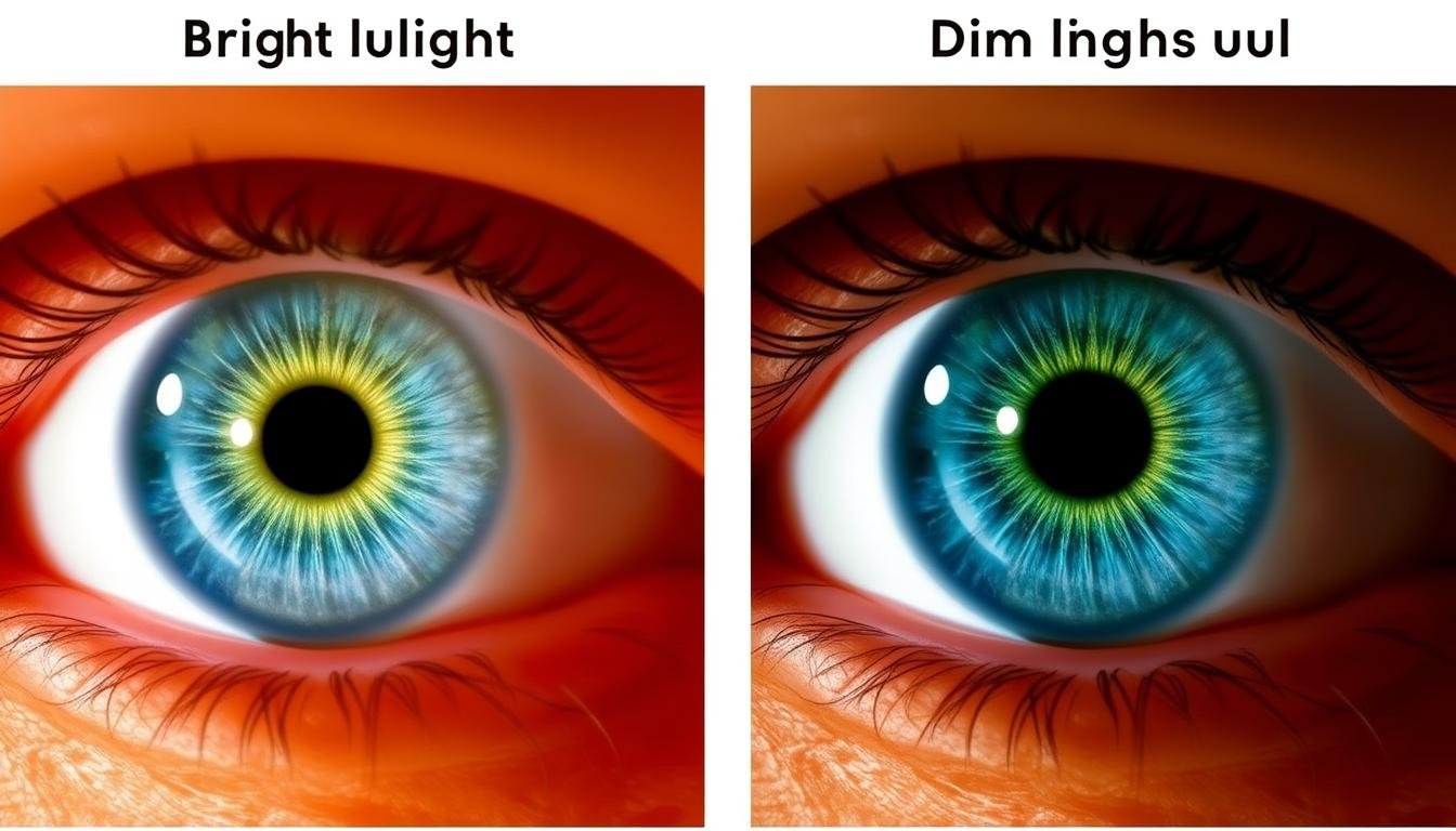
Iris function demonstrated: pupil constriction in bright light (left) versus dilation in dim light (right)
While many people associate the iris primarily with eye color, this remarkable structure performs several critical functions that are essential for vision and eye health.
Light Regulation: The Iris’s Primary Role
The most important function of the iris is controlling the amount of light that enters the eye. It accomplishes this through two sets of muscles:
- Sphincter pupillae – A circular muscle that contracts to constrict the pupil in bright conditions
- Dilator pupillae – Radial muscles that expand the pupil in dim conditions
This dynamic adjustment helps optimize vision across varying light conditions. In bright environments, pupil constriction prevents the retina from being overwhelmed by excessive light. In dim settings, pupil dilation allows maximum light collection for better visibility.

Illustration of iris muscles: sphincter pupillae (circular) and dilator pupillae (radial)
Additional Functions of the Iris
Protection UV
The melanin pigment in the iris helps absorb ultraviolet radiation, providing a protective function for the internal structures of the eye. People with lighter iris colors have less melanin and may be more susceptible to light-related eye damage.
Depth of Field Enhancement
When you focus on nearby objects, your pupils constrict in a process called the accommodative pupillary response. This increases your depth of field (the range of distances that appear in focus), improving visual clarity for close-up tasks.
Aesthetic and Identification Functions
Medical Term Iris: Beyond its physiological roles, the iris provides each person with a unique eye color and pattern. Iris patterns are so distinctive that they’re used in biometric identification systems. The diversity of iris colors—from brown, blue, and green to hazel, gray, and amber—results from variations in melanin concentration and distribution within the iris stroma.
Protect Your Vision
Regular eye examinations can detect early signs of eye conditions before they affect your vision.
Find an Eye Care Specialist
Iris-Related Conditions and Disorders

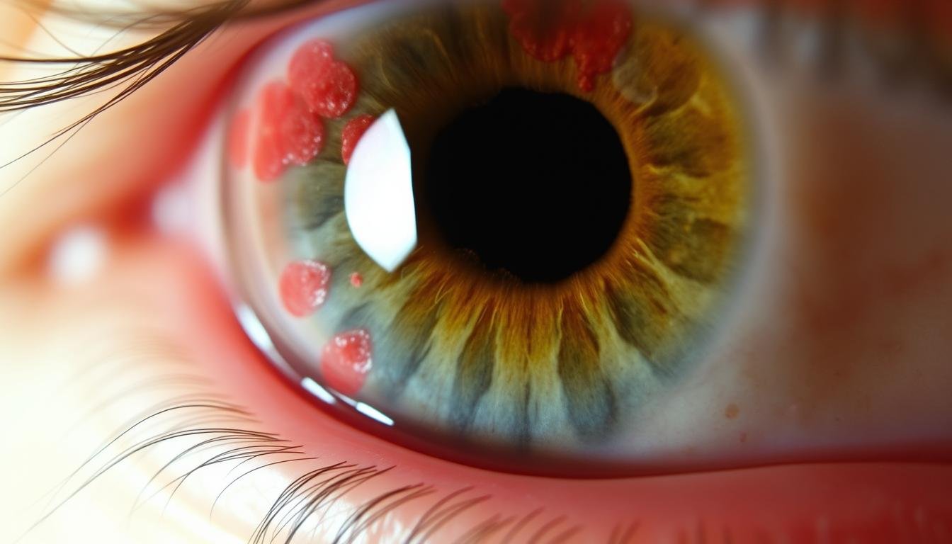
Clinical presentation of acute iritis showing inflammation and irregular pupil
Several conditions can affect the iris, ranging from inflammatory disorders to congenital abnormalities. Understanding these conditions is important for early detection and treatment.
Inflammatory Conditions
IRORS (uvéite précédente)
Iritis is inflammation of the iris and anterior chamber of the eye. It can cause pain, light sensitivity, blurred vision, and a small, irregular pupil. Acute iritis often develops suddenly and may be associated with autoimmune disorders, infections, or trauma. According to a study published in the Journal of Ophthalmology, iritis affects approximately 12 per 100,000 people annually.

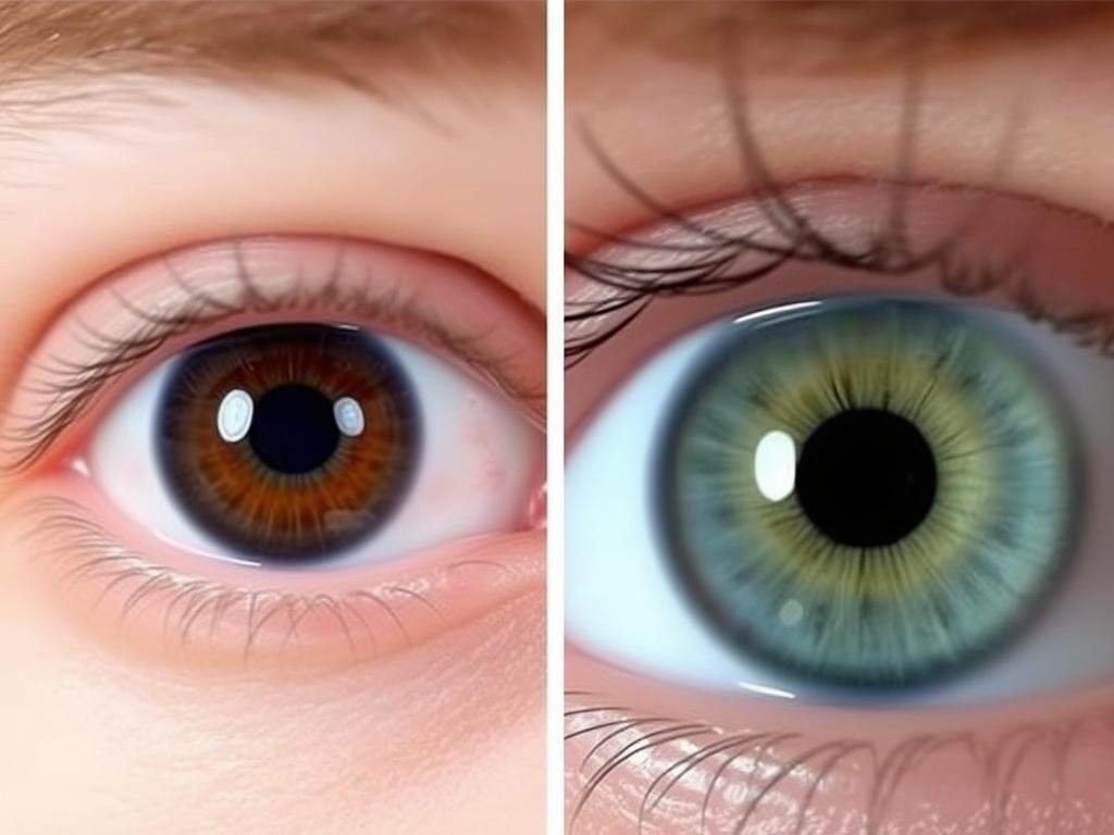
Complete heterochromia: different colored irises in the same individual
Pigmentation Disorders
Hétérochromie
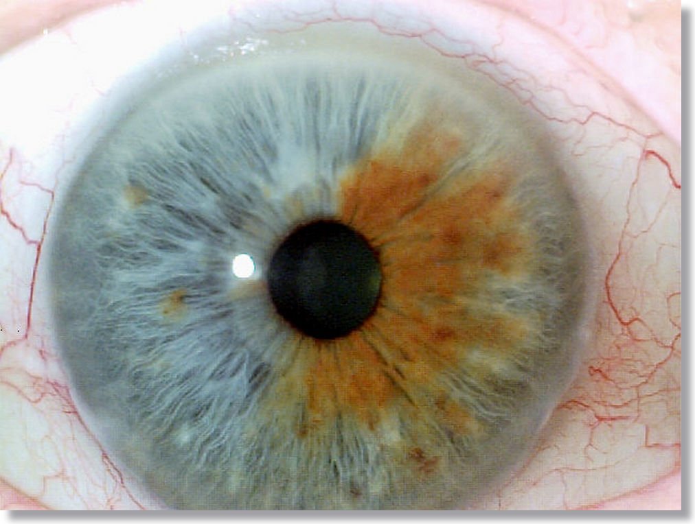
Heterochromia is a condition where a person has different colored irises or different colors within the same iris. Complete heterochromia (heterochromia iridis) involves two differently colored eyes, while partial heterochromia (heterochromia iridium) involves different colors within the same iris. This condition can be congenital or acquired due to injury, inflammation, or certain medications.
Albinisme oculaire
Ocular albinism affects the pigmentation of the eyes without necessarily affecting skin and hair color. People with this condition typically have very light-colored irises that appear translucent due to reduced melanin. This can cause increased light sensitivity and reduced visual acuity. Research in the American Journal of Ophthalmology indicates that ocular albinism affects approximately 1 in 60,000 males.
Structural Abnormalities
Pré-américain
Aniridia is a rare congenital condition characterized by the partial or complete absence of the iris. People with aniridia typically have poor vision, light sensitivity, and are at increased risk for other eye problems like glaucoma and cataracts. The condition affects approximately 1 in 50,000 to 100,000 people worldwide.
Colombma
An iris coloboma appears as a keyhole-shaped pupil due to incomplete formation of the iris during embryonic development. This gap in the iris structure can cause increased light sensitivity and blurred vision. Colobomas may occur as isolated findings or as part of genetic syndromes.

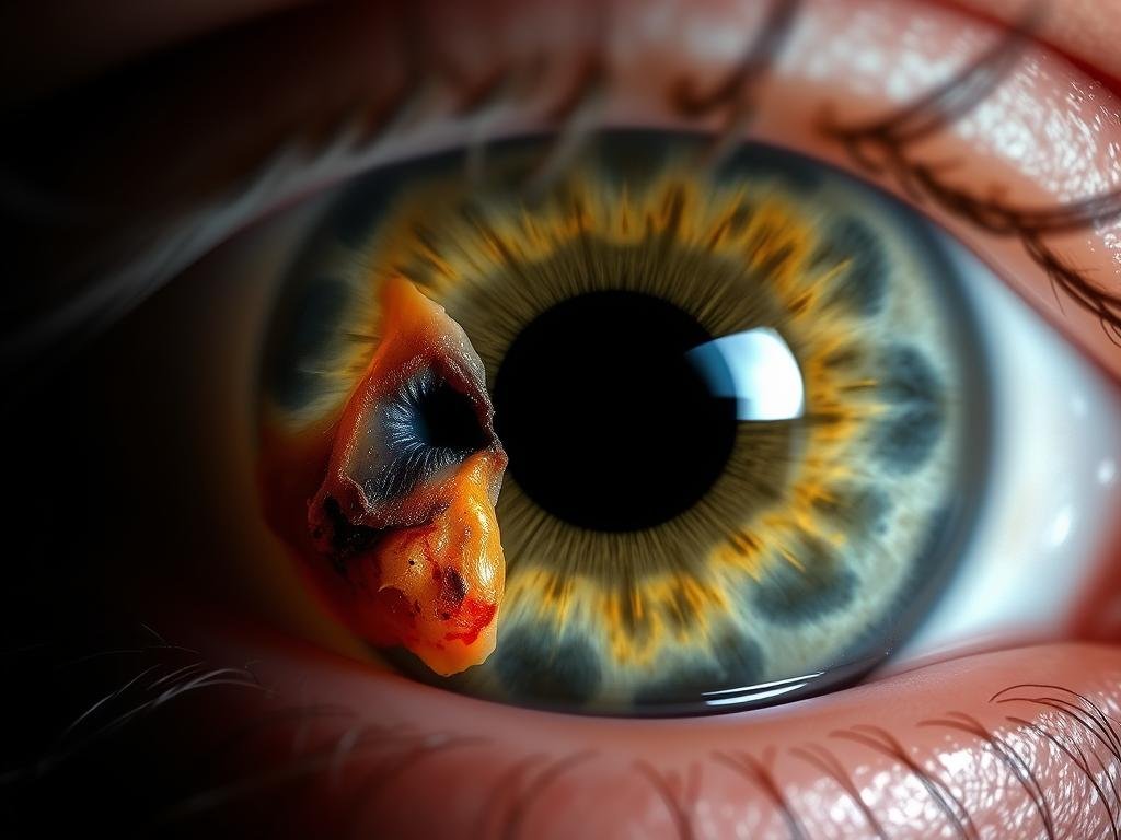
Traumatic injury to the iris showing structural damage and irregular pupil
Traumatic Injuries
Le iris can be damaged by blunt or penetrating eye injuries. Common traumatic iris conditions include:
- Iridodialysis – Separation of the iris from its attachment at the ciliary body
- Traumatic mydriasis – Permanent pupil dilation due to sphincter muscle damage
- Iris tears – Ruptures in the iris tissue that may affect pupillary function
According to a study in the British Journal of Ophthalmology, approximately 15% of all eye injuries involve damage to the iris structure.
Experiencing Eye Problems?
If you’re experiencing changes in vision, eye pain, or unusual iris appearance, consult with an eye specialist promptly.
Learn More About Iris Conditions
Diagnostic Importance of the Iris

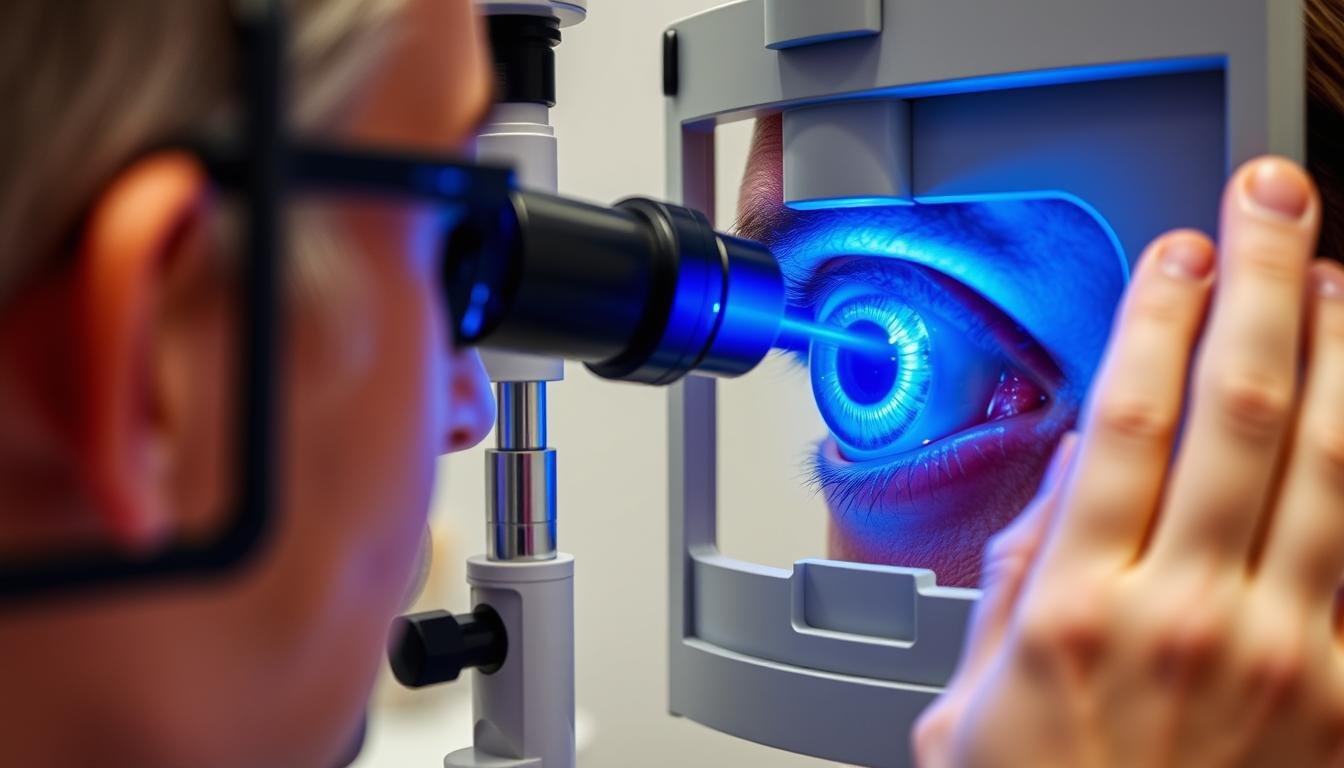
Ophthalmologist examining the iris using a slit lamp biomicroscope
The iris plays a significant role in various diagnostic assessments, providing valuable information about ocular and systemic health.
Iris Examination in Routine Eye Care
During a comprehensive eye examination, eye care professionals carefully assess the iris for:
- Color and pigmentation patterns
- Structural integrity and symmetry
- Pupillary responses to light and accommodation
- Signs of inflammation or abnormal blood vessels
- Presence of nodules, cysts, or other growths
These observations can reveal early signs of ocular conditions or systemic diseases that manifest in the iris. For example, certain inflammatory patterns may suggest autoimmune disorders, while specific iris changes might indicate diabetes or hypertension.
Neurological Assessment
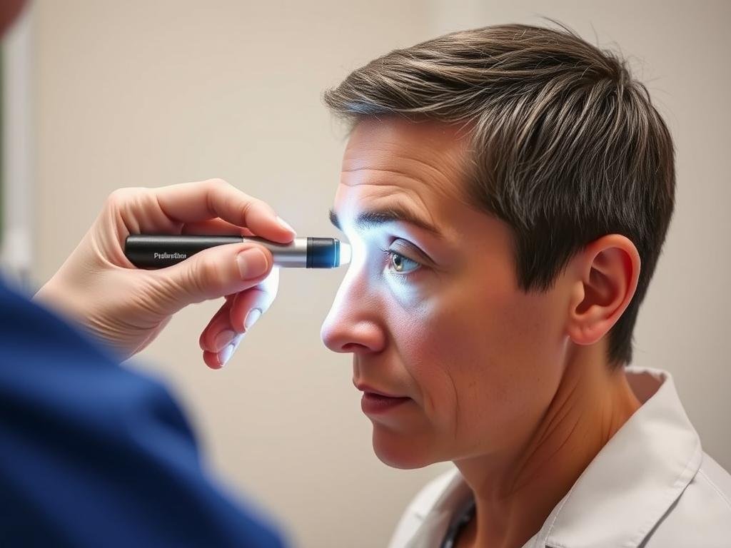
The pupillary light reflex—controlled by the iris muscles—is a critical component of neurological examinations. Abnormal pupillary responses can indicate various neurological conditions, including:
- Brain injuries or increased intracranial pressure
- Oculomotor nerve (CN III) dysfunction
- Horner’s syndrome
- Effects of certain medications or substances

Pupillary light reflex testing – an important neurological assessment of iris function
According to research published in the Journal of Neurology, pupillary assessment has a sensitivity of over 90% for detecting certain types of brain injuries when performed correctly.
Des questions fréquemment posées sur l'iris
Can iris color change throughout life?
While most people’s iris color stabilizes by age 3, some gradual changes can occur throughout life. Babies, particularly those of Caucasian descent, are often born with blue or gray irises that may darken during early childhood as melanin production increases. In adults, certain medications (like prostaglandin analogs used for glaucoma) can gradually darken iris color. Additionally, some eye conditions or injuries may alter iris appearance. However, dramatic spontaneous changes in adult iris color are rare and should be evaluated by an eye care professional.
What causes heterochromia (different colored irises)?
Medical Term Iris: Heterochromia can be congenital (present from birth) or acquired later in life. Congenital heterochromia is typically caused by genetic mutations affecting melanin production or distribution in the iris. It may occur as an isolated finding or as part of syndromes like Waardenburg syndrome. Acquired heterochromia can result from eye injuries, certain medications, inflammation (like chronic iritis), or conditions such as Horner’s syndrome. While often harmless, new-onset heterochromia should be evaluated by an eye specialist to rule out underlying conditions.
How does iritis (iris inflammation) affect vision?
Iritis can significantly impact vision through several mechanisms. The inflammation causes light sensitivity (photophobia) and pain, making it difficult to function in normal lighting conditions. Inflammatory cells in the anterior chamber can cloud vision, while inflammatory debris can settle on the lens, causing blurring. The inflamed iris may also stick to the lens (posterior synechiae), affecting pupil function and potentially leading to complications like glaucoma or cataracts. Prompt treatment with anti-inflammatory medications is essential to prevent permanent vision damage.
Les dommages à l’iris peuvent-ils être réparés ?
Treatment options for iris damage depend on the type and extent of injury. Minor iris tears may heal without intervention, while more significant damage might require surgical repair. Iridoplasty can reshape the iris, while iris prostheses or implants can address larger defects. However, not all iris damage can be fully restored to pre-injury appearance or function. Modern surgical techniques have improved outcomes for traumatic iris injuries, but prevention through protective eyewear remains the best approach. Any iris injury should receive prompt medical attention to minimize complications.
What does it mean if my pupil doesn’t respond normally to light?
Medical Term Iris: Abnormal pupillary responses can indicate various conditions affecting the iris muscles, nerves, or brain. A sluggish or absent pupillary light reflex might suggest oculomotor nerve damage, while unequal pupil sizes (anisocoria) could indicate Horner’s syndrome, Adie’s tonic pupil, or medication effects. In emergency settings, abnormal pupillary responses may signal increased intracranial pressure or brain injury. Certain medications and eye drops can also affect pupil reactivity. Any new or unexplained change in pupillary response warrants prompt medical evaluation, as it could represent anything from a benign condition to a medical emergency.
Understanding Your Iris for Better Eye Health
The iris is much more than just the colored part of your eye—it’s a complex structure that plays a crucial role in vision and can provide important insights into your overall health. From regulating light entry to creating your unique eye color, the iris performs several vital functions that contribute to clear vision and eye protection.
By understanding normal iris anatomy and function, you can better recognize potential problems and seek appropriate care when needed. Regular comprehensive eye examinations are essential for monitoring iris health and detecting conditions early, when they’re most treatable.
If you notice any changes in your iris appearance, pupil function, or vision, consult with an eye care professional promptly. With proper care and attention, you can help maintain the health of this remarkable structure and protect your vision for years to come.

