Das menschliche Auge ist ein bemerkenswertes Organ, das es uns ermöglicht, die Welt um uns herum wahrzunehmen. Diese umfassende Untersuchung der Augen erforscht die komplexe Anatomie, Funktion und Pflege dieser lebenswichtigen Sinnesorgane. Von der Hornhaut bis zur Netzhaut: Wenn wir verstehen, wie unsere Augen funktionieren, können wir ihre Komplexität und Bedeutung für unser tägliches Leben erkennen. Tauchen Sie mit uns in die faszinierende Wissenschaft des Sehens ein und entdecken Sie, wie diese kleinen, aber leistungsstarken Organe Licht verarbeiten, um die Bilder zu erzeugen, die wir sehen.Anatomie des menschlichen Auges: Eine detaillierte Untersuchung der Augen

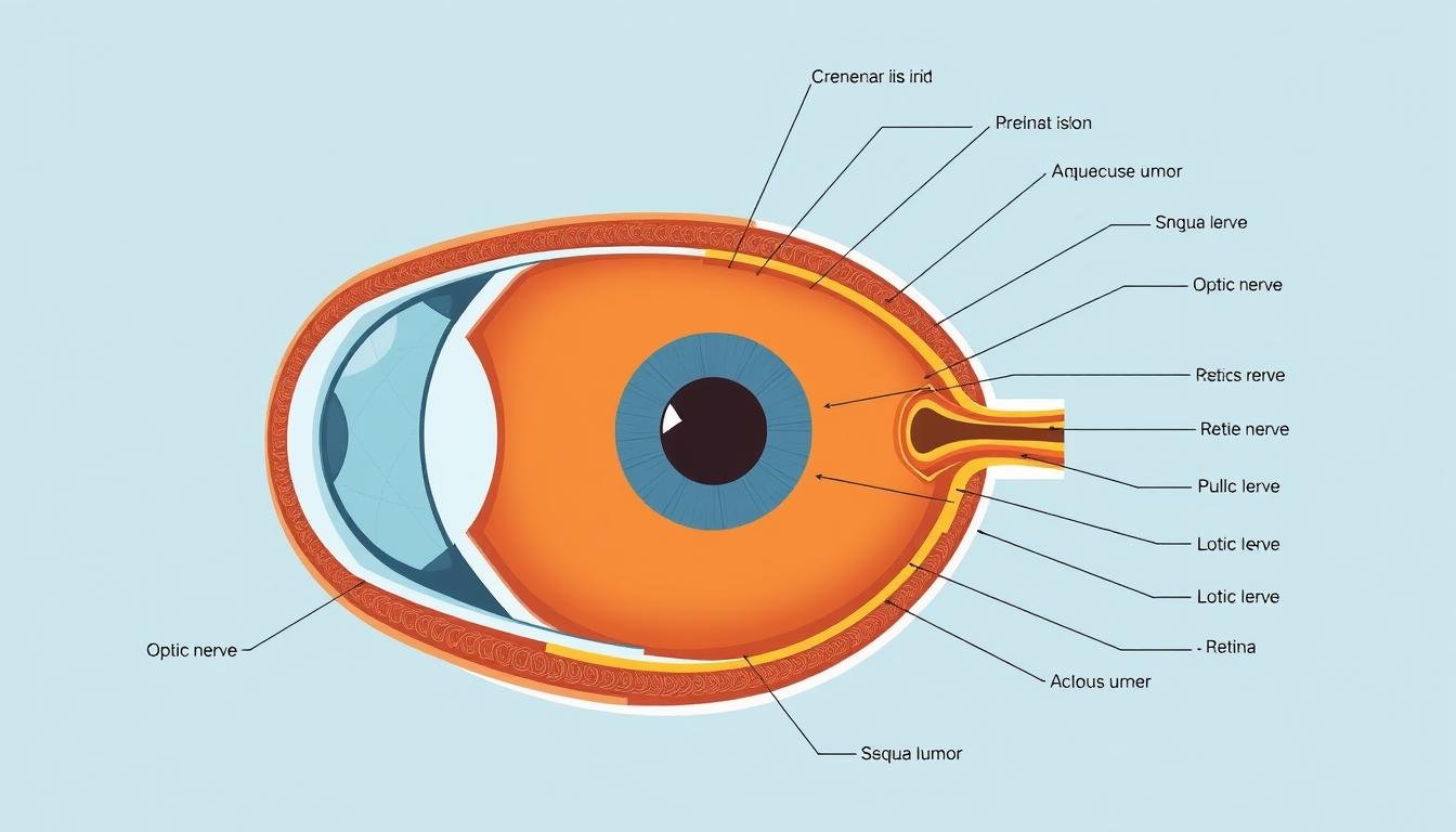
Das Auge ist ein komplexes Organ, das aus mehreren spezialisierten Strukturen besteht, die zusammenarbeiten, um Licht einzufangen und zu verarbeiten. Jede Komponente spielt dabei eine entscheidende Rolle Studium der Augen und Verständnis der Sehmechanik.
Äußere Schicht: Der Schutzschild des Auges
Die äußere Augenschicht besteht aus zwei Hauptstrukturen:
- Hornhaut: Diese klare, kuppelförmige Oberfläche bedeckt die Vorderseite des Auges. Sie fungiert als äußerste Linse des Auges und kontrolliert und fokussiert den Lichteintritt. Die Hornhaut trägt etwa 65–75 % der gesamten Fokussierungsleistung des Auges bei.
- Sklera: Oft genannt “Weiß des Auges,” Dieses robuste, faserige Gewebe schützt die inneren Bestandteile und erhält die Form des Auges. Es bedeckt etwa 83 % der Augenoberfläche.
Mittlere Schicht: Die Gefäßkomponente
Die mittlere Schicht enthält Strukturen, die für die Lichtregulierung und Blutversorgung wichtig sind:
- Iris: Dieser farbige Teil des Auges steuert, wie viel Licht die Netzhaut erreicht, indem er die Größe der Pupille anpasst. Die Iris enthält Pigmente, die die Augenfarbe bestimmen.
- Schüler: Die schwarze kreisförmige Öffnung in der Mitte der Iris, durch die Licht in das Auge eindringen kann. Es dehnt sich bei schwachem Licht aus und verengt sich bei hellem Licht.
- Ziliarkörper: Diese Struktur produziert Kammerwasser und enthält Muskeln, die die Form der Linse zum Fokussieren verändern.
- Aderhaut: Eine Schicht aus Blutgefäßen, die die Netzhaut und andere Teile des Auges mit Sauerstoff und Nährstoffen versorgt.
Innere Schicht: Lichterkennung und -verarbeitung
In der innersten Schicht wird Licht in neuronale Signale umgewandelt:
- Retina: Dieses lichtempfindliche Gewebe kleidet den Augenhintergrund aus. Es enthält Millionen von Fotorezeptoren, die Stäbchen (für das Sehen bei schlechten Lichtverhältnissen) und Zapfen (für das Sehen von Farben und Details) genannt werden.
- Makula: Ein kleiner, spezialisierter Bereich der Netzhaut, der für das zentrale Sehen und die Wahrnehmung feiner Details verantwortlich ist. Die Fovea im Zentrum der Makula bietet die schärfste Sicht.
- Sehnerv: Dieses Bündel aus mehr als einer Million Nervenfasern transportiert visuelle Informationen von der Netzhaut zum Gehirn.
Flüssigkeitssysteme: Erhaltung der Augengesundheit
Zwei wichtige Flüssigkeiten füllen die Augenkammern:
- Wässriger Humor: Eine klare Flüssigkeit, die die Vorderkammer (zwischen Hornhaut und Iris) und die Hinterkammer (zwischen Iris und Linse) füllt. Es liefert Nährstoffe und hält den Augendruck aufrecht.
- Glaskörperhumor: Eine gelartige Substanz, die den großen Hohlraum hinter der Linse ausfüllt und dabei hilft, die Augenform beizubehalten und die Netzhaut an Ort und Stelle zu halten.
Verbessern Sie Ihr Verständnis der Augenanatomie
Laden Sie unser detailliertes Diagramm zur Augenanatomie herunter, um es bei der weiteren Untersuchung der Augen als Referenz zu verwenden. Perfekt für Studenten, medizinisches Fachpersonal oder alle, die sich für Sehwissenschaft interessieren.
Laden Sie das Augenanatomiediagramm herunter
Wie das Sehen funktioniert: Die Wissenschaft hinter dem Sehen

Der Prozess des Sehens ist ein bemerkenswertes Beispiel dafür, wie unser Körper äußere Reize in bedeutungsvolle Informationen umwandelt. Diese Augenstudie offenbart den komplizierten Weg vom Licht zur Wahrnehmung.
Der Weg des Lichts: Von der Welt zur Netzhaut
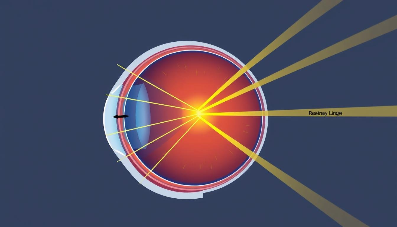
Das Sehen beginnt, wenn Licht von Objekten reflektiert wird und in unsere Augen gelangt:
- Lichteintritt: Lichtstrahlen passieren zunächst die Hornhaut, die das Licht beugt (bricht).
- Schülerordnung: Die Iris passt die Pupillengröße an, um die Lichtmenge zu steuern, die in das Auge eindringt.
- Objektivfokussierung: Die Linse bricht das Licht weiter und passt ihre Form (Akkommodation) an, um Bilder in unterschiedlichen Entfernungen zu fokussieren.
- Glaskörperpassage: Licht wandert durch den Glaskörper und erreicht die Netzhaut.
- Netzhautprojektion: Das Bild wird verkehrt herum und von links nach rechts auf die Netzhaut projiziert.
Fotorezeption: Licht in neuronale Signale umwandeln
Die Netzhaut enthält spezialisierte Zellen, die Licht in elektrische Signale umwandeln:
- Stangen: Ungefähr 120 Millionen Stäbchenzellen sorgen für Schwarzweißsehen und funktionieren auch bei schlechten Lichtverhältnissen gut.
- Kegel: Etwa 6-7 Millionen Zapfenzellen sorgen für Farbsehen und detailliertes Sehen. Es gibt sie in drei Typen: Rot-, Grün- und Blausensitiv.
- Fototransduktion: Wenn Licht auf diese Photorezeptoren trifft, wandelt ein komplexer biochemischer Prozess Lichtenergie in elektrische Signale um.
Neuronale Verarbeitung: Vom Auge zum Gehirn
Die Reise geht weiter, während Signale vom Auge zum Gehirn wandern:
- Signalintegration: Netzhautzellen verarbeiten und integrieren visuelle Informationen, bevor sie sie an das Gehirn senden.
- Sehnervenübertragung: Elektrische Signale wandern durch den Sehnerv, der etwa eine Million Nervenfasern enthält.
- Chiasma opticum: Am Chiasma opticum kreuzen sich Fasern von der nasalen Netzhaut zur gegenüberliegenden Seite des Gehirns, während die Fasern der temporalen Netzhaut auf derselben Seite bleiben.
- Visuelle Cortex-Verarbeitung: Signale erreichen den primären visuellen Kortex im Hinterhauptslappen, wo die grundlegende Verarbeitung stattfindet.
- Höhere visuelle Verarbeitung: Die Informationen werden dann zur komplexen Verarbeitung von Farbe, Bewegung, Tiefe und Objekterkennung an andere Gehirnbereiche weitergeleitet.
Gehirn-Auge-Koordination: Eine Einbahnstraße
Vision ist kein einseitiger Prozess; Gehirn und Augen arbeiten kontinuierlich zusammen:
- Augenbewegungen: Sechs extraokulare Muskeln steuern präzise Augenbewegungen, gesteuert durch Gehirnsignale.
- Binokulares Sehen: Das Gehirn kombiniert leicht unterschiedliche Bilder von jedem Auge, um eine Tiefenwahrnehmung (Stereopsis) zu erzeugen.
- Visuelle Aufmerksamkeit: Das Gehirn weist die Augen an, sich auf bestimmte interessierende Objekte zu konzentrieren.
- Visuelles Gedächtnis: Vergangene Erfahrungen helfen dem Gehirn, das Gesehene zu interpretieren, Lücken zu schließen und vertraute Objekte zu erkennen.
5 überraschende Fakten über das Sehen von Tieren

Fangschreckenkrebs: Der Farbchampion
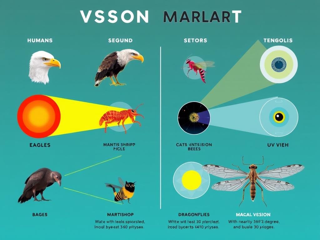
Während das menschliche Auge über drei Arten von Farbrezeptoren (Zapfen) verfügt, verfügt die Fangschreckenkrebse über 16 verschiedene Arten von Fotorezeptoren. Dadurch können sie ultraviolettes, infrarotes und polarisiertes Licht sehen, das Menschen nicht wahrnehmen können. Ihr komplexes visuelles System kann Farben fast augenblicklich verarbeiten, ohne dass eine umfangreiche Gehirnverarbeitung erforderlich ist.
Adler: Teleskope der Natur
Das Sehvermögen von Adlern ist vier- bis achtmal schärfer als das des Menschen. Ihre Netzhaut enthält fünfmal mehr Fotorezeptoren und sie können ultraviolettes Licht sehen. Ein Adler, der in einer Höhe von 1.000 Fuß fliegt, kann ein Kaninchen erkennen, das sich fast eine Meile entfernt bewegt. Diese außergewöhnliche Sehkraft ist auf ihre im Verhältnis zur Kopfgröße größeren Augen und eine höhere Konzentration von Zapfen in ihrer Netzhaut zurückzuführen.
Libellen: Nahezu 360°-Sicht
Libellen besitzen Facettenaugen mit bis zu 30.000 Facetten, von denen jedes als separater Sehrezeptor fungiert. Dies ermöglicht ihnen eine nahezu 360-Grad-Sicht und die Fähigkeit, Bewegungen in einer Entfernung von bis zu 60 Fuß zu erkennen. Ihr Gehirn verarbeitet visuelle Informationen so schnell, dass sie Beute mit einer Genauigkeit von über 95 % verfolgen und abfangen können, selbst wenn sie mit einer Geschwindigkeit von 30 Meilen pro Stunde fliegen.
Chamäleons: Unabhängige Augenkontrolle
Chamäleons können jedes Auge unabhängig voneinander bewegen und haben so die Möglichkeit, gleichzeitig in zwei verschiedene Richtungen zu schauen. Ihre Augen können horizontal um 180 Grad und vertikal um 90 Grad gedreht werden und bieten so ein vollständiges 360-Grad-Sichtfeld, ohne den Kopf bewegen zu müssen. Diese Anpassung hilft ihnen, sowohl Raubtiere als auch Beute zu erkennen, während sie bewegungslos bleiben.
Jakobsmuscheln: Hunderte primitiver Augen
Jakobsmuscheln haben bis zu 200 kleine Augen am Rand ihres Mantels. Im Gegensatz zu unseren Kameraaugen enthält jedes Muschelauge einen konkaven Spiegel aus Kristallen, der Licht auf eine Netzhaut reflektiert. Diese einzigartige Struktur ermöglicht es ihnen, Licht, Dunkelheit und Bewegung zu erkennen, was ihnen hilft, Raubtieren zu entkommen, obwohl sie kein Gehirn haben, wie wir es verstehen.
Diese faszinierenden Anpassungen verdeutlichen die unterschiedliche Art und Weise, wie sich das Sehvermögen bei den verschiedenen Arten entwickelt hat. Während das menschliche Auge bemerkenswert ist, offenbart unsere Untersuchung der Augen im gesamten Tierreich die unglaubliche Vielfalt visueller Systeme, die sich entwickelt haben, um unterschiedlichen Umweltbedürfnissen und Überlebensstrategien gerecht zu werden.
Schützen Sie Ihre wertvolle Vision
Holen Sie sich unseren umfassenden Leitfaden zur Augenpflege mit praktischen Tipps zur Erhaltung gesunder Augen im digitalen Zeitalter, zur Vorbeugung häufiger Erkrankungen und zum Verständnis, wann Sie professionelle Hilfe suchen sollten.
Laden Sie den kostenlosen Leitfaden zur Sehhilfe herunter
Häufige Augenerkrankungen: Sehprobleme verstehen

Da sich unser Verständnis der Augengesundheit verbessert, hat die Untersuchung der Augen zahlreiche Erkrankungen aufgedeckt, die das Sehvermögen beeinträchtigen können. Hier sind einige der häufigsten Störungen und ihre Auswirkungen auf die Sehfunktion:
Brechungsfehler: Wenn der Fokus versagt
Myopie (Kurzsichtigkeit)
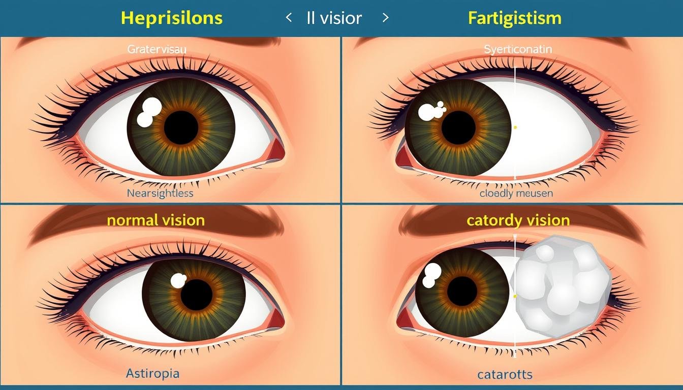
Myopie betrifft etwa 30 % der US-Bevölkerung und tritt auf, wenn der Augapfel zu lang oder die Hornhaut zu stark gekrümmt ist. Dadurch wird das Licht vor der Netzhaut fokussiert und nicht direkt auf diese. Menschen mit Kurzsichtigkeit können Objekte in der Nähe deutlich sehen, entfernte Objekte erscheinen jedoch verschwommen. Laut WHO-Statistiken aus dem Jahr 2023 sind weltweit etwa 2,6 Milliarden Menschen von Kurzsichtigkeit betroffen, und bis 2050 werden voraussichtlich 50 % der Weltbevölkerung davon betroffen sein.
Hyperopie (Weitsichtigkeit)
Das Gegenteil von Myopie, Hyperopie, tritt auf, wenn der Augapfel zu kurz oder die Hornhaut zu flach ist. Das Licht wird hinter der Netzhaut fokussiert, wodurch Objekte in der Nähe verschwommen erscheinen, während entfernte Objekte möglicherweise klarer erscheinen. Ungefähr 5–10 % der Amerikaner leiden unter erheblicher Weitsichtigkeit. Mit zunehmendem Alter macht sich die Erkrankung oft deutlicher bemerkbar, da die Augenlinse an Flexibilität verliert.
Astigmatismus
Diese häufige Erkrankung entsteht durch eine unregelmäßig geformte Hornhaut oder Linse, die dazu führt, dass das Licht auf mehrere Punkte und nicht auf einen einzelnen Punkt auf der Netzhaut fokussiert wird. Dadurch entsteht in allen Entfernungen verschwommenes oder verzerrtes Sehen. Etwa jeder Dritte hat einen gewissen Grad an Astigmatismus, oft zusammen mit Myopie oder Hyperopie.
Presbyopie
Presbyopie ist ein natürlicher Teil des Alterns und beeinträchtigt die Fähigkeit des Auges, sich auf nahe Objekte zu konzentrieren. Typischerweise macht es sich im Alter zwischen 40 und 45 Jahren bemerkbar, wenn die Linse weniger flexibel wird. Laut WHO-Daten aus dem Jahr 2023 sind weltweit schätzungsweise 1,8 Milliarden Menschen von Presbyopie betroffen, darunter 80 % der Erwachsenen über 45.
Altersbedingte Augenkrankheiten: Der Einfluss der Zeit
Katarakte
Beim Grauen Star kommt es zu einer Trübung der Augenlinse, die zu verschwommenem Sehen, verblassten Farben und erhöhter Blendempfindlichkeit führt. Sie sind weltweit die häufigste Ursache für Blindheit. Statistiken der WHO aus dem Jahr 2023 zeigen, dass Katarakte für 51 % der weltweiten Blindheit verantwortlich sind und etwa 94 Millionen Menschen betreffen. Die Erkrankung betrifft vor allem ältere Erwachsene, wobei mehr als die Hälfte der Amerikaner im Alter von 80 Jahren einen Katarakt oder eine Kataraktoperation erleiden.
Altersbedingte Makuladegeneration (AMD)
AMD schädigt die Makula, den zentralen Teil der Netzhaut, der für scharfes, detailliertes Sehen verantwortlich ist. Es kommt zu einem Verlust des zentralen Sehvermögens, während das periphere Sehvermögen intakt bleibt. Laut WHO-Daten aus dem Jahr 2023 sind weltweit 196 Millionen Menschen von AMD betroffen und in Industrieländern die häufigste Ursache für schwere Sehbehinderungen bei Menschen über 60 Jahren.
Andere signifikante Augenerkrankungen
Glaukom
Oft genannt “stiller Dieb des Sehens,” Ein Glaukom schädigt den Sehnerv, typischerweise aufgrund eines erhöhten Augendrucks. Dies führt zu einem allmählichen Verlust des peripheren Sehvermögens, der möglicherweise unbemerkt bleibt, bis ein erheblicher Schaden aufgetreten ist. Der WHO-Bericht 2023 zeigt, dass Glaukom weltweit etwa 76 Millionen Menschen betrifft und die zweithäufigste Ursache für Blindheit weltweit ist.
Diabetische Retinopathie
Diese Erkrankung ist eine Komplikation von Diabetes und schädigt die Blutgefäße in der Netzhaut. Im Anfangsstadium kann es zu keinen Symptomen kommen, im weiteren Verlauf kann es jedoch zu Sehverlust kommen. Nach Angaben der WHO aus dem Jahr 2023 sind weltweit etwa 146 Millionen Menschen von diabetischer Retinopathie betroffen und die häufigste Ursache für Sehverlust bei Erwachsenen im erwerbsfähigen Alter in Industrieländern.
Globale Sehbehinderungsstatistik (WHO 2023): Ungefähr 2,2 Milliarden Menschen weltweit haben eine Sehbehinderung, wobei mindestens 1 Milliarde Fälle vermeidbar sind oder noch behandelt werden müssen. Regionen mit niedrigem und mittlerem Einkommen tragen etwa 90 % der weltweiten Belastung durch Sehbehinderung.
Neueste Forschungsdurchbrüche in der Augenheilkunde

Das Gebiet der Augenheilkunde schreitet weiterhin rasant voran und neue Entdeckungen verbessern unser Verständnis und die Behandlung von Augenerkrankungen. Jüngste Durchbrüche in der Augenforschung haben aufregende Möglichkeiten für die Sehhilfe eröffnet:
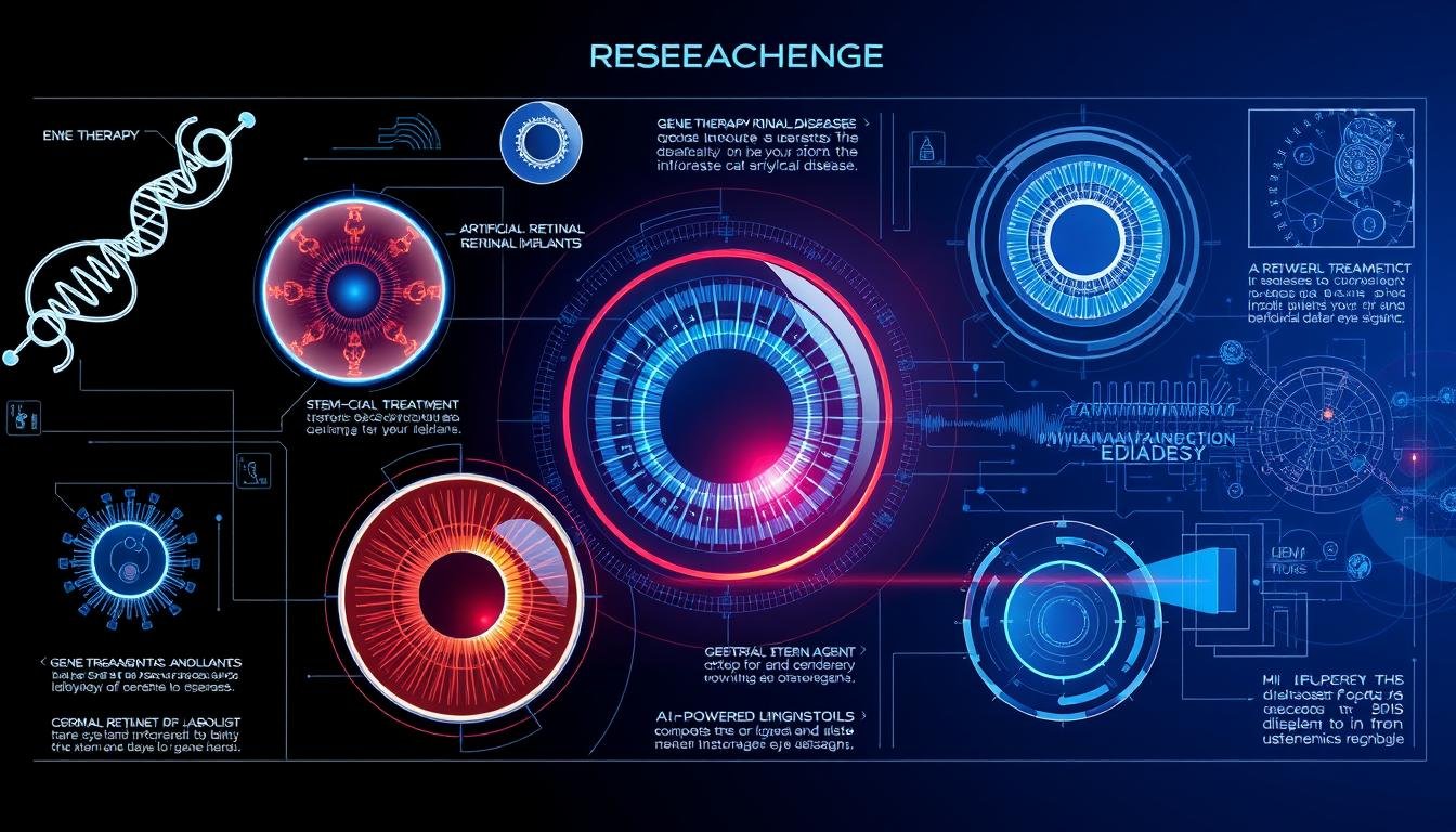
Gentherapie bei Netzhauterkrankungen
Wissenschaftler haben bemerkenswerte Fortschritte bei der Behandlung erblicher Netzhauterkrankungen durch Gentherapie gemacht. Im Jahr 2023 setzten Forscher die CRISPR-Cas9-Genbearbeitung erfolgreich ein, um Mutationen zu korrigieren, die für Retinitis pigmentosa in menschlichen Stammzellen verantwortlich sind. Dieser Ansatz ist vielversprechend für die Behandlung verschiedener genetischer Augenerkrankungen, indem funktionsfähige Gene bereitgestellt werden, um defekte Gene zu ersetzen.
“Die Gentherapie stellt einen Paradigmenwechsel in der Art und Weise dar, wie wir mit bisher unbehandelbaren erblichen Augenkrankheiten umgehen.” sagt Dr. Elena Markov, Direktorin für Netzhautforschung am International Vision Institute. “Wir sind jetzt in der Lage, die zugrunde liegenden genetischen Ursachen anzugehen, anstatt nur die Symptome zu behandeln.”
Künstliche Intelligenz in der Diagnose
KI-Algorithmen haben eine bemerkenswerte Genauigkeit bei der Erkennung von Augenerkrankungen anhand von Netzhautbildern gezeigt. Aktuelle Studien zeigen, dass Deep-Learning-Systeme diabetische Retinopathie, Glaukom und AMD mit einer Genauigkeit von über 95 % erkennen können und dabei manchmal menschliche Spezialisten übertreffen. Diese Instrumente sind besonders wertvoll für das Screening in unterversorgten Gebieten mit eingeschränktem Zugang zu Augenärzten.
Die Integration von KI in tragbare Bildgebungsgeräte hat Möglichkeiten für Fernuntersuchungen und frühere Eingriffe geschaffen und möglicherweise Millionen Menschen vor vermeidbarem Sehverlust bewahrt.
Bionische Augen und visuelle Prothetik
Fortschritte in der Bioelektronik haben zur Entwicklung von Netzhautimplantaten geführt, die Menschen mit bestimmten Formen der Blindheit teilweise das Sehvermögen wiederherstellen können. Die neueste Generation dieser Geräte bietet Bilder mit höherer Auflösung und eine bessere Integration in das visuelle Verarbeitungssystem des Gehirns.
Forscher erforschen außerdem Gehirn-Computer-Schnittstellen, die geschädigte Augen vollständig umgehen und visuelle Informationen direkt an den visuellen Kortex des Gehirns senden. Erste klinische Studien haben vielversprechende Ergebnisse für diesen Ansatz gezeigt.
Regenerative Medizin
Die Stammzelltherapie hat ein bemerkenswertes Potenzial zur Behandlung von Hornhautschäden und bestimmten Netzhauterkrankungen gezeigt. Wissenschaftlern ist es gelungen, Miniaturnetzhäute (Organoide) aus Stammzellen zu züchten und damit wertvolle Modelle für die Untersuchung der Augenentwicklung und -krankheit bereitzustellen.
“Die Fähigkeit, funktionelles Netzhautgewebe im Labor zu züchten, hat die Art und Weise, wie wir Augenkrankheiten untersuchen und mögliche Behandlungen testen, revolutioniert.” erklärt Dr. James Chen, Professor für Regenerative Ophthalmologie an der Pacific Medical University. “Dieser Ansatz ermöglicht es uns, personalisierte Modelle unter Verwendung der eigenen Zellen eines Patienten zu erstellen.”
Innovationen bei der Arzneimittelverabreichung
Neuartige Arzneimittelverabreichungssysteme verändern die Behandlung chronischer Augenerkrankungen. Implantate mit verlängerter Freisetzung können nun Medikamente für bis zu drei Jahre abgeben, wodurch die Notwendigkeit häufiger Injektionen verringert wird. Forscher haben auch Kontaktlinsen entwickelt, die Medikamente langsam freisetzen und so die Einhaltung und Wirksamkeit der Behandlung bei Erkrankungen wie Glaukom und trockenem Auge verbessern.
Telemedizin und Fernüberwachung
Die Integration der Smartphone-Technologie mit speziellen Aufsätzen hat Augenuntersuchungen und -überwachungen aus der Ferne ermöglicht. Patienten können jetzt bestimmte Sehtests zu Hause durchführen und die Ergebnisse an ihren Augenarzt übermitteln. Dieser Ansatz hat sich als besonders wertvoll für die Behandlung chronischer Erkrankungen und die Verbesserung des Zugangs zur Gesundheitsversorgung in ländlichen Gebieten erwiesen.
Bleiben Sie über Fortschritte in der Sehwissenschaft auf dem Laufenden
Abonnieren Sie unser vierteljährliches Vision Science Update, um die neuesten Forschungsergebnisse, Behandlungsinnovationen und Empfehlungen zur Augenpflege direkt in Ihren Posteingang zu erhalten.
Abonnieren Sie Vision-Updates
Praktische Augenpflege-Tipps für den Lebensstil des digitalen Zeitalters

In der heutigen digitalen Welt stehen unsere Augen vor beispiellosen Herausforderungen. Längere Bildschirmzeit, künstliche Beleuchtung und der Lebensstil in Innenräumen wirken sich alle auf die Augengesundheit aus. Basierend auf den neuesten Studien zur Augen- und Sehwissenschaft finden Sie hier praktische Empfehlungen zur Aufrechterhaltung einer optimalen Augengesundheit:
Die 20-20-20-Regel: Digitale Augenschutzprävention
Bis zu 65 % der Computernutzer sind von digitaler Augenbelastung betroffen. Bekämpfen Sie dies mit der einfachen 20-20-20-Regel:
- Alle 20 Minuten der Bildschirmzeit
- Schauen Sie sich etwas in einer Entfernung von 20 Fuß an
- Mindestens 20 Sekunden lang
Diese Übung reduziert die Ermüdung der Augenmuskulatur und trägt dazu bei, die Fokussierflexibilität aufrechtzuerhalten. Erwägen Sie die Verwendung einer Timer-App, um sich daran zu erinnern, bis es zur Gewohnheit wird.
Optimale Bildschirmergonomie
Die richtige Positionierung digitaler Geräte reduziert die Belastung der Augen erheblich:
- Positionieren Sie Ihren Monitor ungefähr Armlänge entfernt (20-24 Zoll)
- Der obere Rand des Bildschirms sollte sich auf Höhe oder etwas darunter befinden Augenhöhe
- Bildschirm anpassen Helligkeit passend zu Ihrer Umgebung
- Verwenden matte Bildschirmfilter Blendung zu reduzieren
- Erwägen Sie die Verwendung Nachtmodus oder Blaulichtfilter am Abend
Ernährung für die Augengesundheit
Bestimmte Nährstoffe sind besonders förderlich für die Erhaltung gesunder Augen:
Essentielle Nährstoffe
- Lutein und Zeaxanthin: Diese Carotinoide kommen in Blattgemüse vor und schützen die Makula vor Schäden durch blaues Licht
- Omega-3-Fettsäuren: Sie sind in Fisch, Leinsamen und Walnüssen enthalten, unterstützen die Tränenproduktion und reduzieren Entzündungen
- Vitamin A: Entscheidend für die Nachtsicht und die Gesundheit der Hornhaut, enthalten in Orangengemüse und Leber
- Vitamin C: Ein Antioxidans, das hilft, die Blutgefäße im Auge zu erhalten, reichlich vorhanden in Zitrusfrüchten und Beeren
- Vitamin E: Schützt Zellen vor oxidativen Schäden, die in Nüssen, Samen und Pflanzenölen vorkommen
- Zink: Wichtig für die Gesundheit der Netzhaut und das Nachtsichtvermögen, enthalten in Austern, Rindfleisch und Hülsenfrüchten
Flüssigkeitszufuhr und Augengesundheit
Die richtige Flüssigkeitszufuhr ist für die Tränenproduktion und den allgemeinen Augenkomfort unerlässlich. Ziel ist es:
- Trinken Sie zumindest 8-10 Gläser Wasser täglich
- Limit Alkohol und Koffein, was dehydrierend sein kann
- Verwenden Luftbefeuchter in trockenen Umgebungen
- In Betracht ziehen künstliche Tränen ohne Konservierungsstoffe wenn bei Ihnen Symptome eines trockenen Auges auftreten
Schutzmaßnahmen
Der Schutz Ihrer Augen vor Umweltgefahren ist von entscheidender Bedeutung:
- UV-Schutz: Tragen Sie eine Sonnenbrille, die auch an bewölkten Tagen 99–100 % der UVA- und UVB-Strahlung blockiert
- Schutzbrillen: Tragen Sie bei gefährlichen Tätigkeiten, beim Sport oder bei der Arbeit mit Werkzeugen einen geeigneten Augenschutz
- Kontaktlinsenhygiene: Befolgen Sie die ordnungsgemäßen Reinigungs- und Austauschpläne, um Infektionen vorzubeugen
- Händehygiene: Vermeiden Sie es, Ihre Augen mit ungewaschenen Händen zu berühren oder zu reiben
Regelmäßige Augenuntersuchungen
Umfassende Augenuntersuchungen sind für die Erhaltung der Augengesundheit unerlässlich:
- Erwachsene 18–60: Alle 2 Jahre (häufiger, wenn Risikofaktoren vorliegen)
- Erwachsene 61+: Jährlich
- Kontaktlinsenträger: Jährlich
- Menschen mit Diabetes oder Bluthochdruck: Jährlich oder wie von Ihrem Arzt empfohlen
Denken Sie daran, dass sich viele schwere Augenerkrankungen ohne frühe Symptome entwickeln. Regelmäßige Untersuchungen können Probleme erkennen, bevor sie Ihr Sehvermögen beeinträchtigen.
Häufig gestellte Fragen zum Thema Sehvermögen und Augengesundheit

Wie sehen Augen Farben?
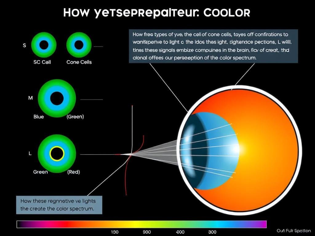
Das Farbsehen beginnt mit spezialisierten Zellen in der Netzhaut, den sogenannten Zapfen. Menschen haben typischerweise drei Arten von Zapfen, die jeweils auf unterschiedliche Lichtwellenlängen empfindlich reagieren:
- S-Kegel (kurze Wellenlänge): Am empfindlichsten gegenüber blauem Licht (420-440 nm)
- M-Zapfen (mittlere Wellenlänge): Am empfindlichsten gegenüber grünem Licht (534-545 nm)
- L-Zapfen (lange Wellenlänge): Am empfindlichsten gegenüber rotem Licht (564-580 nm)
Wenn Licht in das Auge gelangt, werden diese Zapfen je nach vorhandener Wellenlänge unterschiedlich stark stimuliert. Das Gehirn interpretiert diese unterschiedlichen Muster der Zapfenaktivierung als bestimmte Farben. Wenn beispielsweise alle drei Zapfenarten gleichermaßen stimuliert werden, nehmen wir weißes Licht wahr. Das Fehlen einer Stimulation wird schwarz angezeigt.
Dieses trichromatische System ermöglicht es dem Menschen, etwa 1 Million verschiedene Farben zu unterscheiden. Einige Tiere, wie Fangschreckenkrebse, verfügen über mehr Arten von Photorezeptoren und können ultraviolettes und polarisiertes Licht wahrnehmen, das Menschen nicht sehen können.
Können Augenübungen das Sehvermögen verbessern?
Die Wirksamkeit von Augenübungen hängt davon ab, welchen Aspekt des Sehvermögens Sie verbessern möchten:
- Bei Fehlsichtigkeiten (Kurzsichtigkeit, Weitsichtigkeit, Astigmatismus): Wissenschaftliche Erkenntnisse stützen nicht die Behauptung, dass Augenübungen diese Beschwerden korrigieren können. Diese Sehprobleme resultieren aus der physischen Form des Auges oder der Alterung der Linse, die durch Übungen nicht verändert werden kann.
- Bei bestimmten binokularen Sehproblemen: Eine Sehtherapie, ein strukturiertes Programm visueller Aktivitäten, das von Augenärzten verordnet wird, kann bei bestimmten Erkrankungen wie Konvergenzinsuffizienz (Schwierigkeit, beide Augen bei der Naharbeit zusammen zu fokussieren) und bestimmten Arten von Strabismus (Augenfehlstellung) wirksam sein.
- Bei digitaler Augenbelastung: Einfache Übungen wie die 20-20-20-Regel, Konzentrationsübungen und bewusstes Blinzeln können dabei helfen, die Symptome einer digitalen Augenbelastung zu lindern, ändern aber nichts an Ihrer Sehstärke.
Wenn Sie Sehprobleme haben, wenden Sie sich für die richtige Diagnose und Behandlung am besten an einen Optiker oder Augenarzt, anstatt sich ausschließlich auf Augenübungen zu verlassen.
Warum erweitern und verengen sich die Pupillen?
Die Pupillengröße wird durch zwei Muskelgruppen in der Iris gesteuert:
- Pupillensphinkter: Zirkuläre Muskeln, die sich zusammenziehen, um die Pupille zu verkleinern (Konstriktion)
- Pupillendilatator: Radiale Muskeln, die sich zusammenziehen, um die Pupille zu vergrößern (Dilatation)
Die Größe der Schüler ändert sich aus mehreren Gründen:
- Lichtregulierung: Bei hellem Licht verengen sich die Pupillen, um den Lichteinfall in das Auge zu verringern. Bei schwachem Licht dehnen sie sich aus, um mehr Licht hereinzulassen.
- Fokuseinstellung: Während der Nahfokussierung (Akkommodation) verengen sich die Pupillen leicht, um die Schärfentiefe zu erhöhen und die Bildschärfe zu verbessern.
- Emotionale Reaktionen: Die Pupillen weiten sich bei emotionaler Erregung, sei es positiv (Aufregung) oder negativ (Angst).
- Arzneimittelwirkungen: Bestimmte Medikamente können eine Pupillenerweiterung (Mydriasis) oder eine Pupillenverengung (Miosis) verursachen.
- Medizinische Bedingungen: Abnormale Pupillenreaktionen können auf neurologische Probleme oder Augenverletzungen hinweisen.
Dieses dynamische Reaktionssystem trägt dazu bei, die Sicht bei unterschiedlichen Lichtverhältnissen und Aktivitäten zu optimieren.
Wichtige Erkenntnisse aus unserer Augenstudie

Anatomie und Funktion des Auges verstehen
- Das Auge ist ein komplexes Organ mit mehreren Schichten und Strukturen, die harmonisch zusammenarbeiten, um das Sehen zu ermöglichen
- Licht durchläuft mehrere Strukturen (Hornhaut, Kammerwasser, Linse, Glaskörper), bevor es die Netzhaut erreicht
- Photorezeptoren in der Netzhaut wandeln Licht in elektrische Signale um, die über den Sehnerv zum Gehirn gelangen
- Das Gehirn verarbeitet diese Signale, um die Bilder zu erzeugen, die wir wahrnehmen
Schutz des Sehvermögens in der modernen Welt
- Die Nutzung digitaler Geräte erfordert regelmäßige Pausen nach der 20-20-20-Regel
- Die richtige Ernährung, Flüssigkeitszufuhr und UV-Schutz sind für die langfristige Augengesundheit unerlässlich
- Regelmäßige umfassende Augenuntersuchungen können Probleme erkennen, bevor sie das Sehvermögen beeinträchtigen
- Viele schwerwiegende Augenerkrankungen entwickeln sich ohne frühe Symptome, weshalb die Vorbeugung von entscheidender Bedeutung ist
Die Untersuchung der Augen verrät nicht nur, wie wir die Welt sehen, sondern bietet auch einen Einblick in unsere allgemeine Gesundheit. Während die Sehwissenschaft voranschreitet, entwickeln wir weiterhin bessere Möglichkeiten, diesen wertvollen Sinn zu schützen, zu bewahren und möglicherweise wiederherzustellen.
— Dr. James Chen, Professor für Regenerative Ophthalmologie
Unsere Augen sind bemerkenswerte Organe, die angemessene Pflege und Aufmerksamkeit verdienen. Indem wir verstehen, wie sie funktionieren, und vorbeugende Maßnahmen ergreifen, können wir ein Leben lang ein gesundes Sehvermögen bewahren. Egal, ob Sie sich für die Wissenschaft hinter dem Sehen interessieren oder nach praktischen Möglichkeiten zur Pflege Ihrer Augen suchen, diese umfassende Augenstudie bietet eine Grundlage für die Wertschätzung und den Schutz eines unserer wertvollsten Sinne.
Vervollständigen Sie Ihre Ausbildung in Vision Science
Laden Sie unseren kompletten Satz Augenanatomiediagramme, Leitfäden zur Sehpflege und aktuelle Forschungszusammenfassungen in einem praktischen Paket herunter.
Holen Sie sich das komplette Vision Science-Paket

