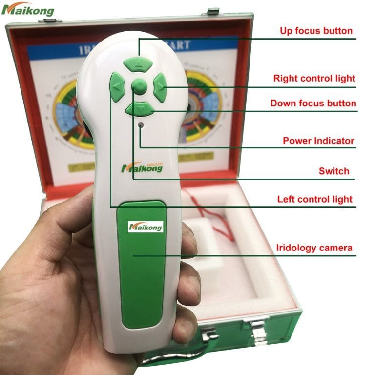
What is 12MP DigitaI Eye Iriscope camera Iridology USB camera?


12MP DigitaI Eye Iriscope camera Iridology USB camera Specifications
1. Résolution haute, Real 2 Magepixel Pictures
2. Fonctionnement facile sans installation de pilote
3. Ensemble de cuir de luxe
12MP DigitaI Eye Iriscope camera Iridology USB camera Instruction:
* Belle apparence et design innovant
* Illuminateur LED autour de l'objectif
* Lentille importée avec couche plaquée
* Capteur CCD haute résolution de 12 mégapixels
* Processeur d'image DSP spécial, stabilisateur d'image optique
* Bouton de capture unique et capture de pause numérique.
* Mise au point réglable pour donner une image claire.
* Balance des blancs et réglage automatique du contraste, filtre de température de couleur
* Fonction de comparaison d'images double
* Mode de capture 3D négatif
* Compatible avec les lentilles cutanées et capillaires.
* Fournissez des images claires et précises.
* Facile à utiliser.
* Résolution maximale : 3264*2448
* OS: Windows XP, Win2000, 2003, Vista, Win7.Win 8
12MP DigitaI Eye Iriscope camera Iridology USB camera Accessories:
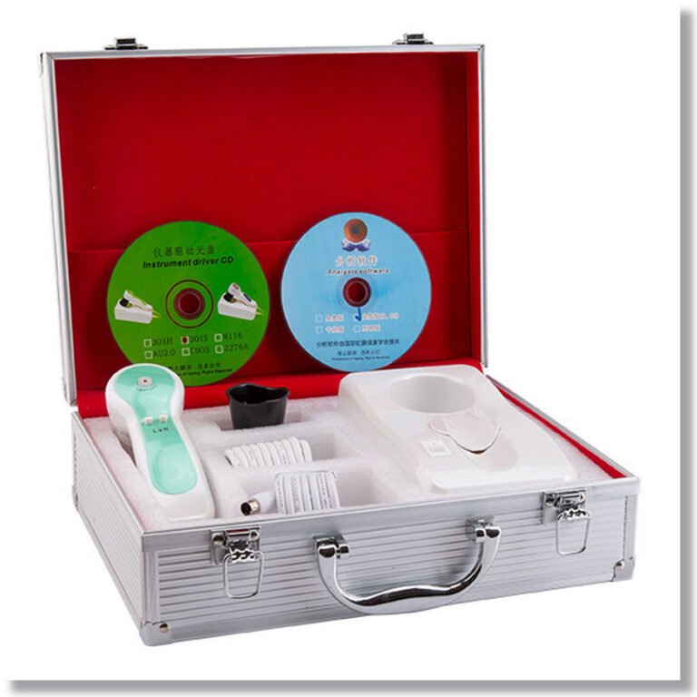
- Combiné X1pc
- 30X Iris Lens X1pc (skin and hair lens/software for option, contact us if need)
- Boîte en cuir PU X1pc
- 1.5 Meter USB Line X1pc
- Couvercle de protection d'objectif X1pc
12MP DigitaI Eye Iriscope camera Iridology USB camera Package:
* Chart d'Iridology x1pc
* Instructions & Garantie X1pc
* CD (logiciel d'analyse du pilote et pro) x1pc


How to use the iriscope camera software?
1 installez le logiciel.
2 connexion clé usb orange avec votre pc.
3 Ouvrez le bureau “Système d'analyse avancé CadiCV version anglaise”
1)utilisez sélectionner “utilisateur”,mot de passe :111111,et cliquez sur : “se connecter”
2)cliquez “outil client”, entrez vos informations client. si ok, cliquez “ajouter”, et cliquez”fermer”
3)cliquez “capturer l'oeil droit”.–cliquez “capturer”,
4) répétition de l'œil gauche Dernière étape.
5) sélectionnez la photo de l'œil (photo de l'œil droit / photo de l'œil gauche)
6)cliquez “analyse”
7)cliquez “définir le paramètre” bouton.
8)cliquez “analyse de l'iris” bouton.
9)analyse
Observation des symptômes et de la couleur de l'iris au choix dans le logiciel au dessus du bouton correspondant aux symptômes et à la nuit.
Par exemple : il y a des fissures sur la photo de l'iris et la couleur est claire.
Sélectionnez le bouton « crack » et le bouton « light », déplacez le curseur sur les fissures de l'iris,
Cliquez avec la souris. Immédiatement rapporté par l'analyse.
Vous et ajoutez votre recommandation ou votre produit pour le client.
ajouter ——? Analyse——–?gauche (Analyse œil gauche)
10 sauvegardes
11 rapport : sélectionnez le nom du rapport —–?Rapport HTML ou rapport privé, sélectionnez le nom du rapport (nom de la date)
12 tirages
13 supprimer le client
14 modifier le client
Comment fonctionne l'iridologie? -Iridology La justification de l'iridologie serait associée aux connexions nerveuses entre l'œil et le cerveau à travers le nerf optique. Cette connexion rend un circuit possible à chaque partie du corps et distingue l'offre nerveuse saine et malsaine. Les fibres d'iris qui réflexes à un organe spécifique qui se trouve dans un état aigu ou chronique seront évidentes par la couleur et la texture.
L'iridologie est l'étude de l'iris associé à la maladie. Les «iris’ de l'œil, est la structure tissulaire la plus complexe de l'anatomie humaine, qui est les terminaisons nerveuses exposées qui composent la partie colorée de l'œil. À partir du moment où nous sommes conçus, l'iris et toutes ses fibres se forment avec le cerveau avant que tout autre organe ne soit développé, faisant des yeux une extension du cerveau doté de milliers de terminaisons nerveuses, de vaisseaux sanguins microscopiques, de muscles et d'autres tissus. L'iris est lié au tissu du corps par le biais du cerveau et du système nerveux. Les fibres nerveuses ramifiées (comme les dendrites) reçoivent leurs impulsions par leurs liens avec le nerf optique et la moelle épinière. Les fibres nerveuses de l'iris réagissent aux changements dans les tissus corporels en manifestant une physiologie réflexe qui correspond à des changements et des emplacements de tissus spécifiques, en termes de modèles de ligne, de structure de fibres et de changements de couleur dans l'iris. Par ce moyen, l'état héréditaire et / ou acquis du corps envoie des réflexes neuronaux aux fibres de l'iris provoquant le changement de groupes de fibres à titre de motifs de ligne, caractéristiques, formes, structures, formes et couleur dans l'iris et c'est ce qu'un iridologue étudie.


How Choosing an iriscope camera?
Essential elements to look for in iriscope cameras
Notre système de fibre optique d'éclairage froid minimise l'irritation du client et donne un éclairage précis pour une exposition optimale et une vraie couleur à chaque fois. Il comprend un ajustement d'intensité variable pour permettre même aux yeux les plus sombres d'afficher chaque détail sans perte de contraste! De plus, nos lumières évitent les élèves pour éviter de mettre des artefacts dans cette zone d'évaluation vitale.
Opération facile. Obtenez une concentration parfaite avec facilité.
L'une des caractéristiques les plus importantes à considérer est l'éclairage. Notre système d'éclairage fixe assure une analyse comparative facile. Option d'éclairage latéral incluse.
Notre concepteur a eu plus de 30 ans d'expérience clinique en tant que ND et iridologue qui apporte avec lui une compréhension approfondie de ce dont les iridologues professionnels ont besoin.
Nous n'utilisons que des caméras de modèle de haute qualité et des matériaux de qualité professionnelle. La plupart de nos systèmes de caméras incluent un logiciel d'analyse de pointe dans le prix.
Tous nos caméras répondent aux critères photographiques de l'Iridology. En fait, nous ouvrons la voie à Iris Photography!
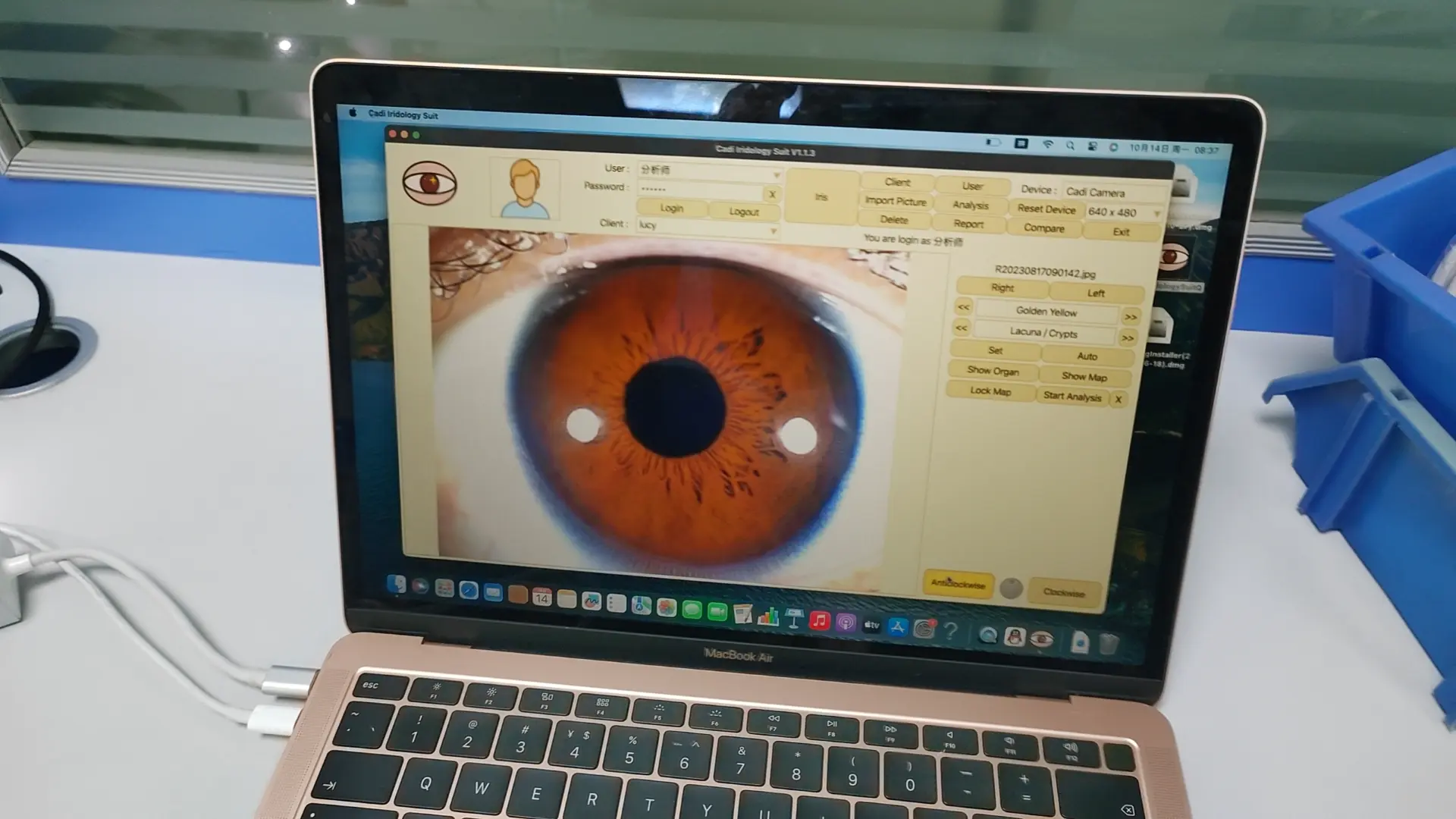
Vidéo de fonctionnement du logiciel
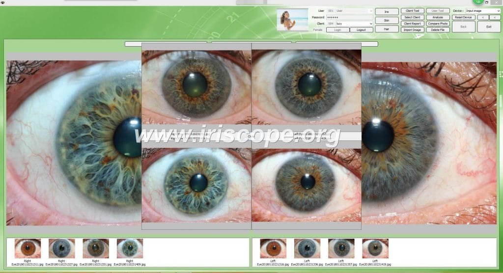
Interface d'exploitation du logiciel
fournisseur de caméra iriscopeWe are offer Top brand Newest iriscope camera manufacturer,We can offer OEM iriscope camera and software services. best factory price.Contact now!


