What is 12MP DigitaI Eye Iriscope camera Iridology USB camera?


12MP DigitaI Eye Iriscope camera Iridology USB camera Specifications
1. Alta risoluzione, REAL 2 Magepixel Immagini
2. Facile operazione senza installazione del driver
3. Pacchetto in pelle di lusso
12MP DigitaI Eye Iriscope camera Iridology USB camera Instruction:
* Bell'aspetto e design innovativo
* Illuminatore a LED intorno all'obiettivo
* Lente importato con strato placcato
* Sensore CCD ad alta risoluzione da 12 mega pixel
* Processore di immagine DSP speciale, stabilizzatore dell'immagine ottica
* Pulsante di acquisizione singolo e cattura di pausa digitale.
* Focus regolabile per dare un'immagine chiara.
* Bilancio bianco e regolazione del contrasto, filtro a temperatura colore
* Funzione di confronto a doppia immagine
* Modalità di acquisizione 3D negativa
* Compatibile con lenti cutanee e lenti per capelli.
* Fornire immagini chiare e accurate.
* Facile da usare.
* Risoluzione massima: 3264* 2448
* OS: Windows XP, Win2000, 2003, Vista, Win7.Win 8
12MP DigitaI Eye Iriscope camera Iridology USB camera Accessories:
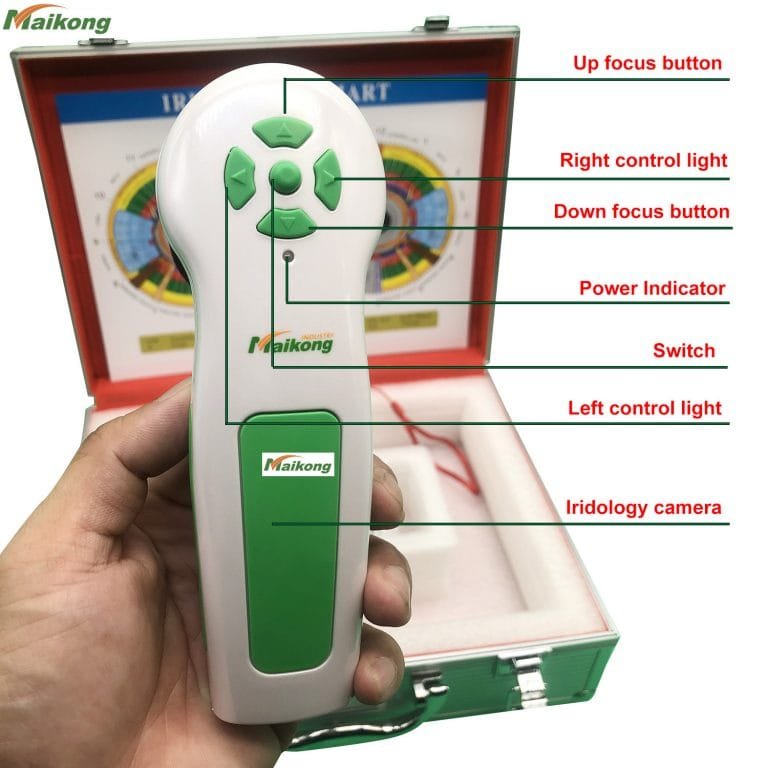
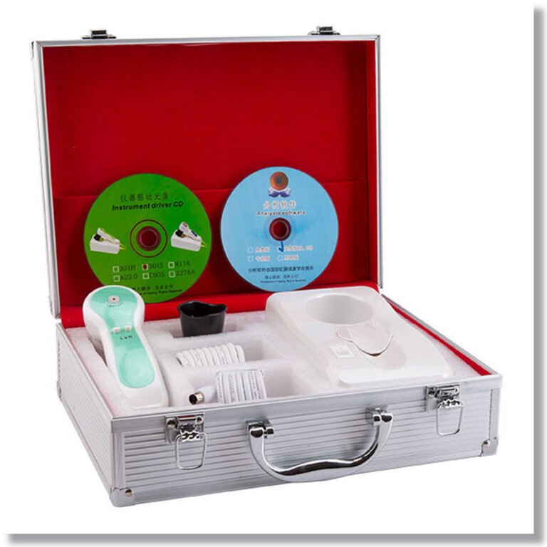
- Handset X1pc
- 30X Iris Lens X1pc (skin and hair lens/software for option, contact us if need)
- Leather PU Box X1pc
- 1.5 Meter USB Line X1pc
- Lens protective cover X1pc
12MP DigitaI Eye Iriscope camera Iridology USB camera Package:
* Grafico IRIDOLOGIA X1PC
* Istruzioni & Garanzia x1pc
* CD (software di analisi driver e pro) X1pc


How to use the iriscope camera software?
1 installare il software.
2 chiavette usb arancioni di connessione al vostro pc.
3 Aprire il desktop “CadiCV Advance Analysis System Versione inglese”
1)usa seleziona “utente”,password:111111,e fare clic su: “login”
2)fare clic “strumento cliente”, inserisci le informazioni del tuo cliente. se ok, clicca “aggiungere”e fare clic su”vicino”
3) fare clic “catturare l'occhio destro”.–clic “catturare”,
4) ripetizione dell'ultimo passaggio con l'occhio sinistro.
5)seleziona l'immagine dell'occhio (immagine dell'occhio destro/immagine dell'occhio sinistro)
6) fare clic “analisi”
7)fare clic “impostare il parametro” pulsante.
8) fare clic “analisi dell'iride” pulsante.
9)analisi
Osservazione dei sintomi e colore dell'iride scelto nel software sopra il pulsante corrispondente sui sintomi e sulla notte.
Ad esempio: c'è una crepa sull'immagine dell'iride e il colore è chiaro.
Selezionare il pulsante "crack" e il pulsante "light", spostare il cursore sulle crepe sull'iride,
Fare clic con il mouse. Subito segnalato dalle analisi.
Tu e aggiungi il tuo consiglio o prodotto per il cliente.
aggiungere ——? Analisi——–?sinistra (Analisi occhio sinistro)
10 salvi
11 rapporto: seleziona il nome del rapporto —–?html report o report privew seleziona il nome del report (nome data)
12 stampa
13 cancella cliente
14 modifica cliente
How iridology work?-IridologyThe rationale behind iridology is believed to be associated with the nerve connections between the eye and brain through the optic nerve. This connection makes a circuit possible to every part of the body and distinguishes between healthy and unhealthy nerve supply. Iris fibers that reflex to a specific organ that is in an acute or chronic state will be evident by color and texture.
Iridology is the study of the iris as associated with disease. The ‘iris’ of the eye, is the most complex tissue structure in the human anatomy, which is the exposed nerve endings that makes up the colored part of the eye. From the time we are conceived the iris and all its fibers are formed along with the brain before any other organ is developed, making the eyes an extension of the brain endowed with thousands of nerve endings, microscopic blood vessels, muscle and other tissues. The iris is connected to the tissue of the body by way of the brain and nervous system. The branched nerve fibers (such as dendrites) receive their impulses by their connections to the optic nerve and spinal cord. Nerve fibers in the iris respond to changes in body tissues by manifesting a reflex physiology that corresponds to specific tissue changes and locations, by way of line patterns, fiber structure and color changes in the iris. By this means the body’s inherited and/or acquired state of health sends neural reflexes to the fibers within the iris causing groups of fibers to change by way of line patterns, characteristics, shapes, structures, forms and color in the iris and that is what an Iridologist studies.


How Choosing an iriscope camera?
Essential elements to look for in iriscope cameras
Il nostro sistema a fibre ottiche con illuminazione fredda riduce al minimo l'irritazione del cliente e fornisce un'illuminazione diurna precisa per un'esposizione ottimale e colori realistici ogni volta. Include la regolazione dell'intensità variabile per consentire anche agli occhi più scuri di visualizzare ogni dettaglio senza perdita di contrasto! Inoltre, le nostre luci evitano le pupille per evitare di inserire artefatti in quest'area di valutazione vitale.
Funzionamento facile. Ottieni una messa a fuoco perfetta con facilità.
Una delle caratteristiche più importanti da considerare è l’illuminazione. Il nostro sistema di illuminazione fissa garantisce una facile analisi comparativa. Opzione di illuminazione laterale inclusa.
Il nostro designer ha più di 30 anni di esperienza clinica come ND e iridologo, portando con sé una profonda comprensione di ciò di cui hanno bisogno gli iridologi professionisti.
Utilizziamo solo fotocamere attuali di alta qualità e materiali di livello professionale. La maggior parte dei nostri sistemi di telecamere includono nel prezzo un software di analisi all'avanguardia.
Tutte le nostre fotocamere soddisfano i criteri fotografici dell'iridologia. In effetti, siamo all'avanguardia nella fotografia dell'iride!
Video sul funzionamento del software
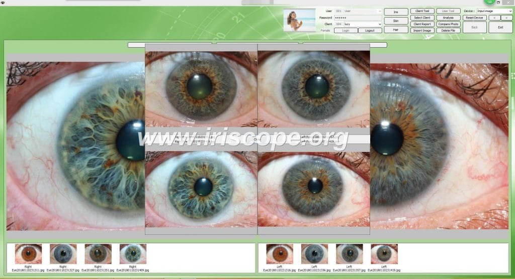
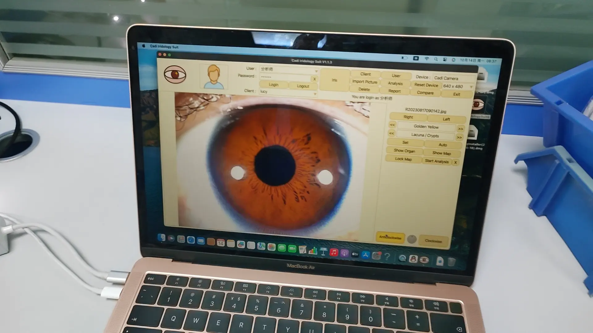
Interfaccia operativa del software
fornitore di fotocamere IriscopeWe are offer Top brand Newest iriscope camera manufacturer,We can offer OEM iriscope camera and software services. best factory price.Contact now!


