The human iris contains remarkable details that may reveal insights about your overall health. Modern iridology combines traditional chart analysis with advanced camera technology to create a comprehensive approach to iris examination. This guide explores how ارڊولوجي چارٽس and specialized cameras work together, providing practitioners and health enthusiasts with powerful tools for iris analysis.ڇا آهي هڪ Iridology چارٽ؟

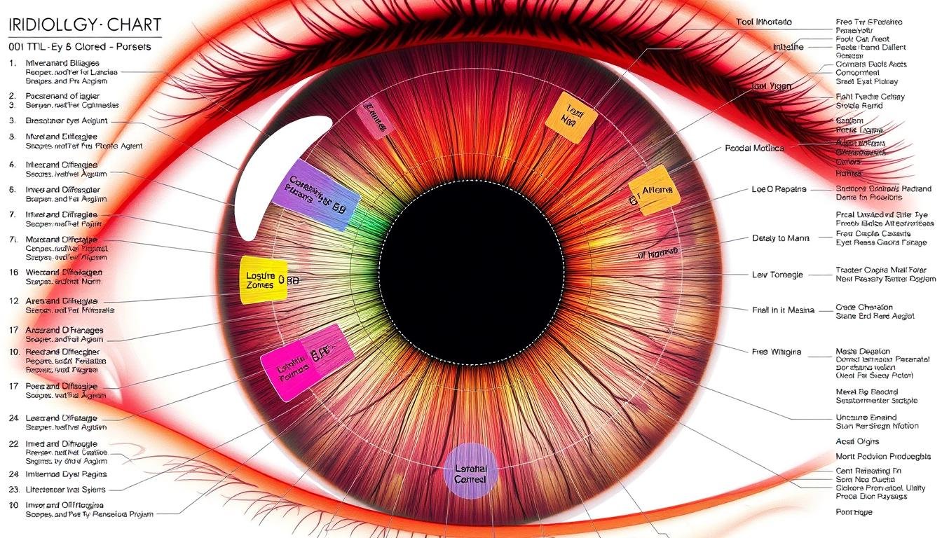
A standard ارڊولوجيٽر چارٽ mapping different zones of the iris to body systems
هڪ ارڊولوجيٽر چارٽ serves as a detailed map that divides the iris—the colored part of your eye—into multiple zones, each corresponding to different organs and systems within the body. These charts are fundamental tools used by iridologists to interpret patterns, colors, and markings in the iris that may indicate health conditions or predispositions.
رواجي ارڊولوجي چارٽس typically display the iris divided into approximately 80-90 zones, creating a complex map that practitioners use as a reference guide during examinations. The right and left irises are mapped differently, with each representing different aspects of the body’s systems.
Key Zones in a Standard Iridology چارٽ
Right Iris Mapping
- اپر علائقو: دماغ، سر، ۽ سينوس
- Right quadrants: Lungs, lymphatic system
- Middle zone: Liver, gallbladder, pancreas
- هيٺيون علائقو: اندريون، پيدائشي عضوو
- Inner ring: Digestive system and stomach
Left Iris Mapping
- Upper region: Cerebral circulation, mental function
- Left quadrants: Heart, spleen
- Middle zone: Kidneys, adrenals
- Lower region: Lymphatic system, elimination
- Inner ring: Autonomic nervous system
The concentric arrangement of these zones follows the body’s natural organization, with areas closer to the pupil typically representing internal organs and digestive system, while outer zones often correspond to skin, muscles, and extremities.
The Role of Iridology Cameras in Analyzing Iridology چارٽس

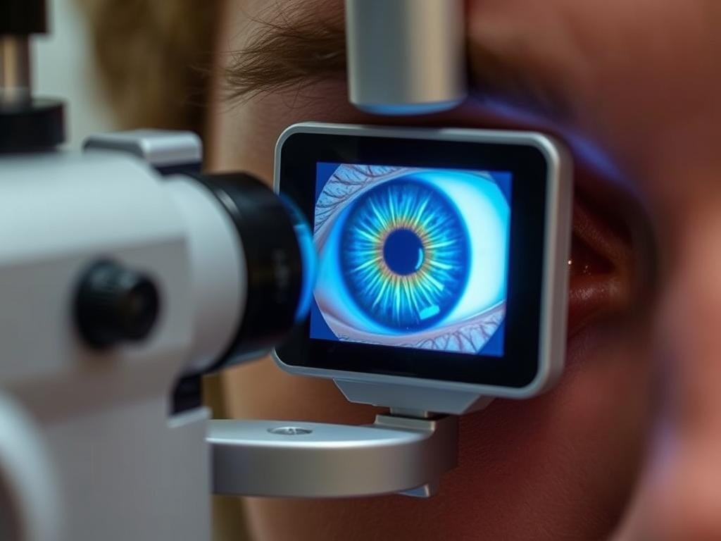
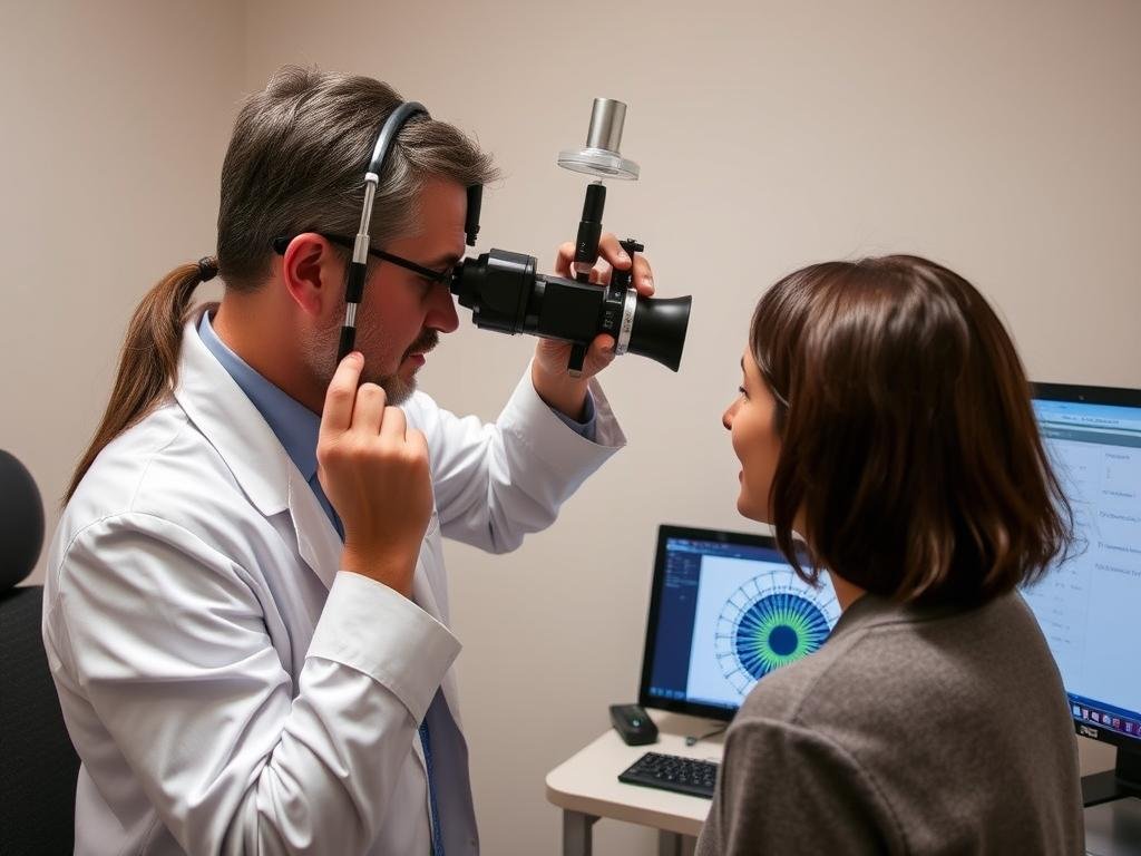
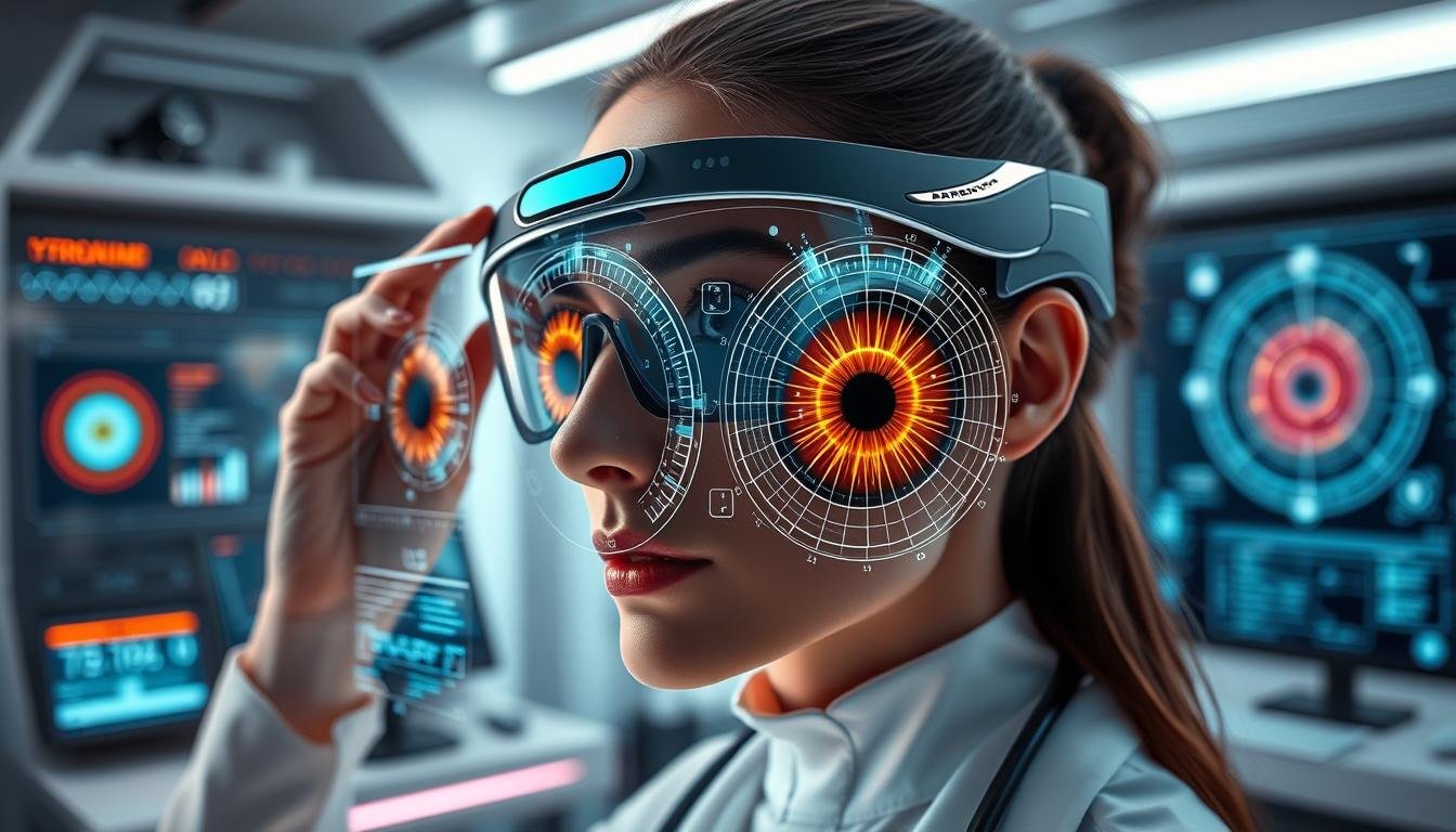
Modern iridology camera capturing high-resolution iris details
While traditional iridology relied on direct observation and simple magnifying tools, modern practice has been revolutionized by specialized iridology cameras. These devices capture high-resolution images of the iris, allowing for detailed analysis when compared against standard ارڊولوجي چارٽس.
Advanced Features of Modern Iridology Cameras
- High-resolution imaging (typically 15+ megapixels)
- Specialized lighting to reveal subtle iris details
- Magnification capabilities up to 40x
- Digital storage of iris images for comparison over time
- Software integration with digital ارڊولوجي چارٽس
These cameras eliminate much of the subjectivity involved in traditional iridology by providing consistent, detailed images that can be analyzed methodically against standardized charts. The technology allows practitioners to identify minute details that might be missed through direct observation alone.
پنھنجي Iridology جي مشق کي وڌايو
Discover how our advanced iridology camera systems can transform your practice with precise iris imaging and integrated chart analysis.
Explore Camera Options
ڪئين Iridology چارٽس and Cameras Work Together

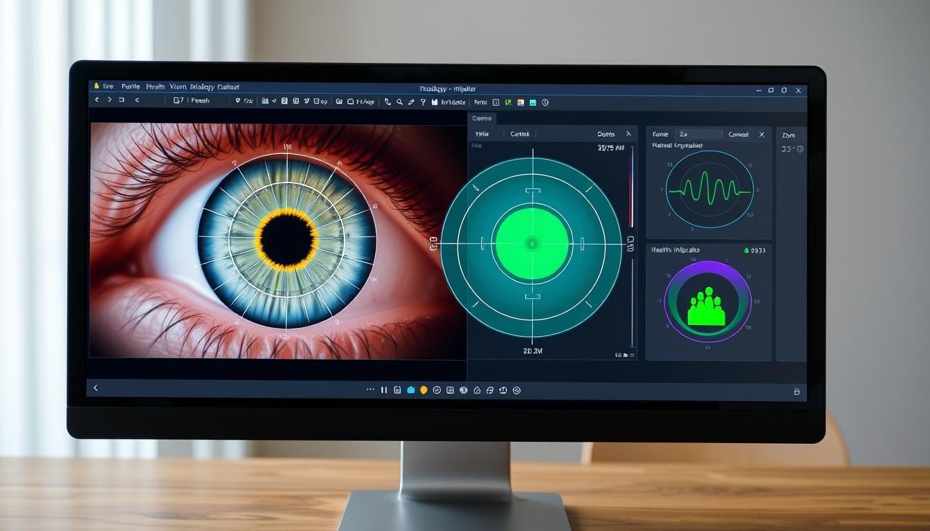
Digital integration of iris photography with ارڊولوجيٽر چارٽ تجزيو
The integration of cameras and charts creates a powerful diagnostic approach that combines technological precision with traditional iridology knowledge. This synergy follows a systematic process:
The Integrated Iridology Process
Step 1: Image Capture
The iridology camera captures high-resolution images of both irises under controlled lighting conditions, ensuring consistent results.
Step 2: Digital Mapping
Software overlays the standard ارڊولوجيٽر چارٽ onto the iris image, aligning the zones precisely with the individual’s unique iris structure.
Step 4: Analysis & تعبير
The practitioner interprets the patterns, colors, and markings according to established iridology principles and the specific zone mappings.
Modern software can enhance this process by automatically detecting certain patterns and providing preliminary analysis based on the ارڊولوجيٽر چارٽ mappings. However, skilled practitioners remain essential for accurate interpretation of these findings.
How to Interpret Iridology چارٽس with Modern Technology

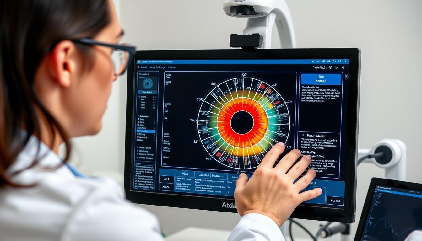
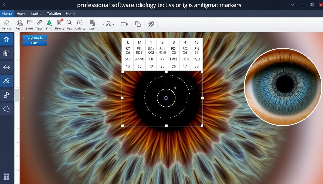
Modern software interface for ارڊولوجيٽر چارٽ تجزيو
Interpreting iris signs requires understanding both the ارڊولوجيٽر چارٽ mappings and the significance of various markings. Modern technology enhances this process through several key approaches:
Key Indicators in Iris Analysis
| آئرس جي نشاني | ظهور | Traditional Interpretation | How Technology Enhances Detection |
| Lacuna | Closed, darkened areas | Potential inherent weakness in the corresponding organ | Digital contrast enhancement reveals subtle lacunae |
| Radii Solaris | ٻاھران نڪرندڙ ڳالھائيندڙ لڪيرون | Toxic conditions in related systems | High-resolution imaging captures fine radii invisible to naked eye |
| پگمينٽيشن | Colored spots or patches | Chemical deposits or drug accumulation | Color analysis software identifies subtle pigment variations |
| Lymphatic Rosary | White cloud-like ring around iris | Lymphatic system congestion | Automated measurement of ring density and completeness |
| نروس رِنگس | Circular lines resembling tree rings | Nervous tension or stress response | Pattern recognition algorithms count and measure ring patterns |
Modern software can track changes in these indicators over time, creating a visual record of potential health changes that would be difficult to monitor through traditional observation alone.
Master Iridology Interpretation
Join our comprehensive online course to learn professional iris analysis using modern chart techniques and camera technology.
Enroll in Training Program
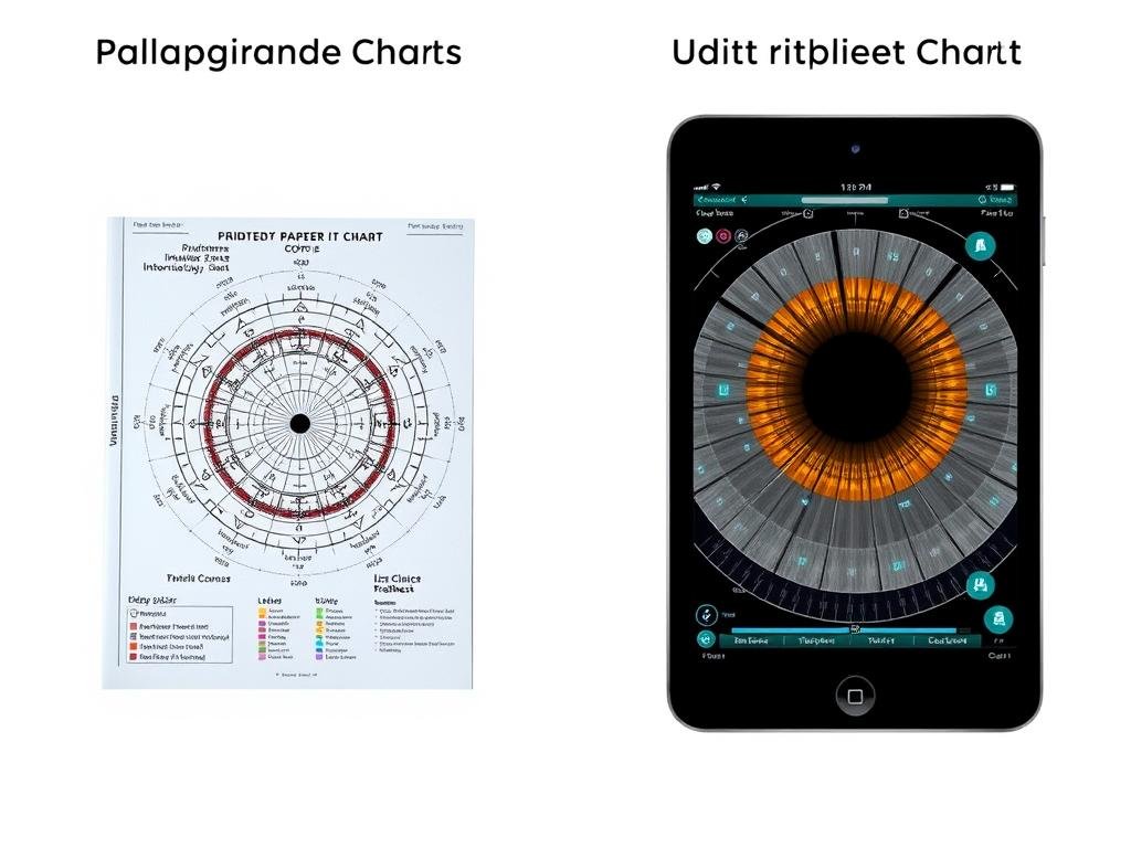
Digital vs. Traditional Iridology چارٽس

Evolution from traditional to digital ارڊولوجي چارٽس
Digital Chart Advantages
- Interactive zooming for detailed examination
- Customizable overlays for different analysis approaches
- Integration with patient records and history
- Automatic alignment with iris photographs
- Regular updates based on latest research
- درست تجزيو لاء ماپ جا اوزار
Traditional Chart Limitations
- Static representation with no interactive features
- Limited detail at different magnification levels
- Manual alignment with actual iris is challenging
- No integration with patient history
- Difficult to update with new findings
- Subjective interpretation without measurement tools
The transition to digital ارڊولوجي چارٽس represents a significant advancement in the field, allowing for more precise analysis and better integration with modern healthcare approaches. However, understanding the traditional chart foundations remains essential for proper interpretation.
Case Studies: Successful Applications of Camera and Iridology چارٽ تجز مبارڪ

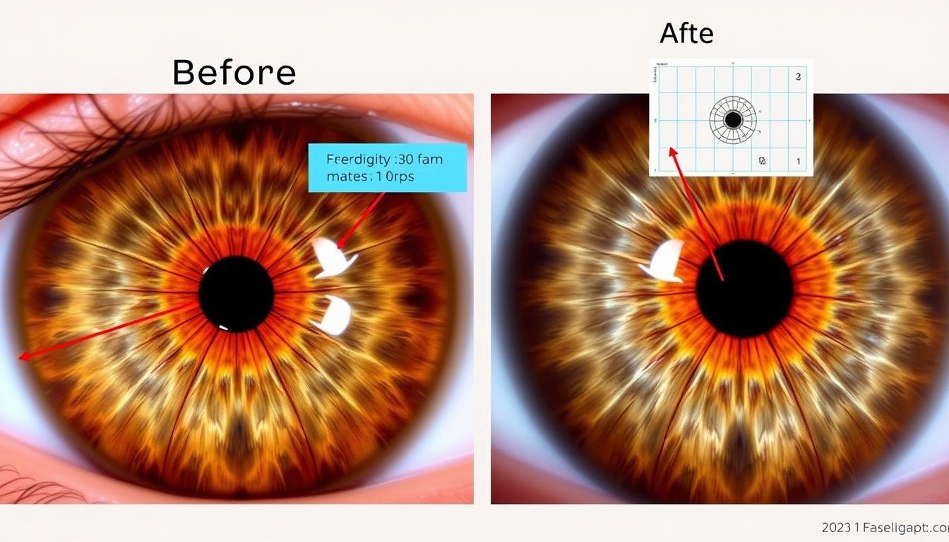
Before and after iris analysis showing changes detected through ارڊولوجيٽر چارٽ mapping
ڪيس جو مطالعو 1: هاضمي سسٽم جو جائزو
A 42-year-old patient with chronic digestive complaints underwent iris analysis using high-resolution camera imaging and digital ارڊولوجيٽر چارٽ mapping. The analysis revealed distinct markings in the zones corresponding to the small intestine and pancreas.
The practitioner recommended dietary modifications and specific digestive support based on these findings. Follow-up imaging three months later showed subtle changes in the iris patterns, coinciding with the patient’s reported improvement in symptoms.
Case Study 2: Stress Response Monitoring
A stress management program utilized iridology camera imaging to track changes in nerve rings (circular contraction furrows) in participants’ irises. Using standardized ارڊولوجيٽر چارٽ analysis, practitioners documented changes in these indicators over an 8-week stress reduction program.
Digital analysis software quantified reductions in nerve ring prominence that correlated with participants’ self-reported stress levels, providing visual feedback on program effectiveness.
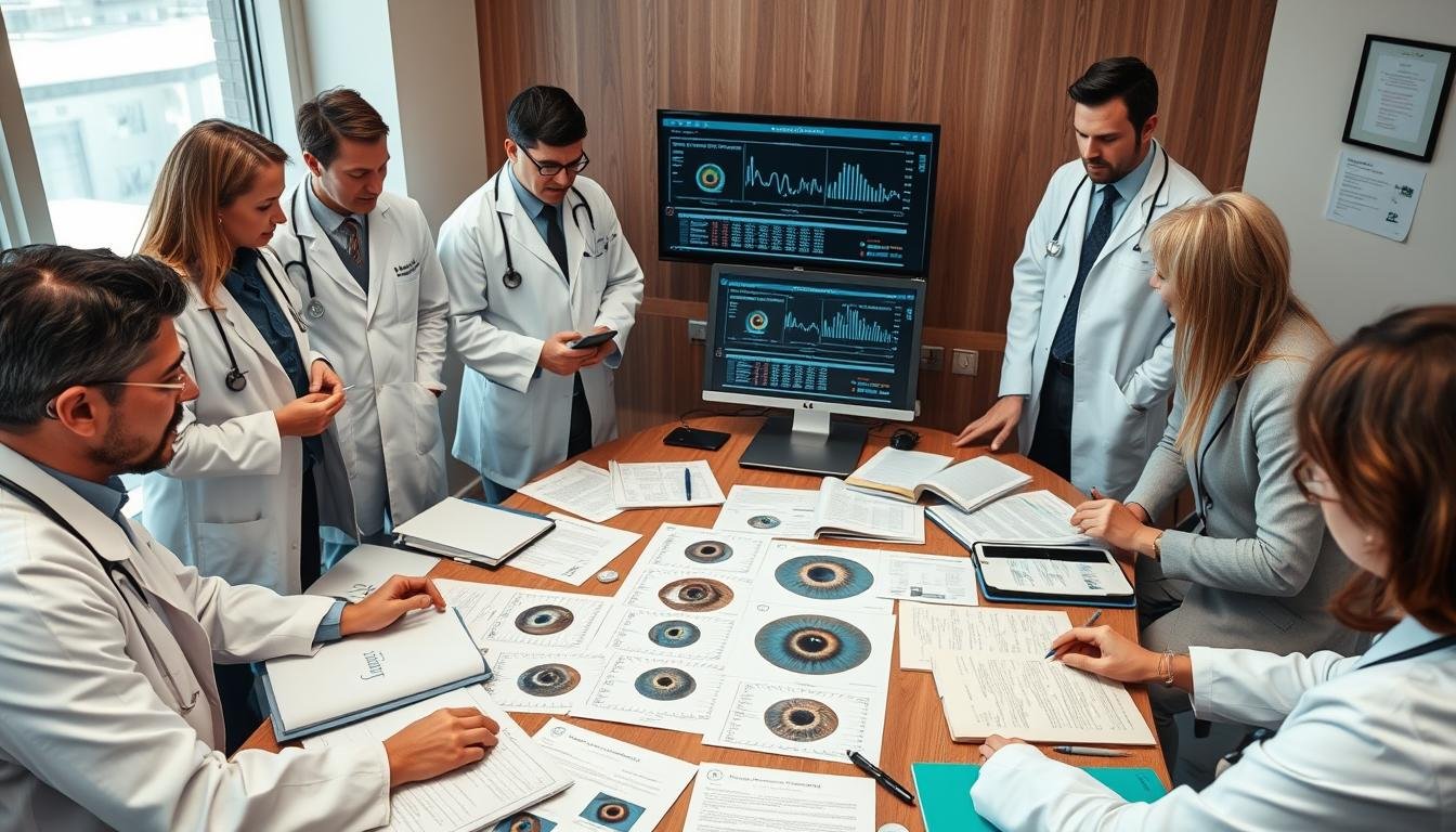
These case studies demonstrate how the combination of advanced camera technology and ارڊولوجيٽر چارٽ analysis can provide visual documentation of potential health changes over time. While not diagnostic in a conventional medical sense, these approaches offer complementary perspectives on health monitoring.
Best Practices for Using Iridology Cameras with Iridology چارٽس

Proper technique for iris photography ensures accurate ارڊولوجيٽر چارٽ تجزيو
Technical Considerations for Optimal Results
Camera Setup
- Use consistent lighting (preferably LED with color temperature 5500-6500K)
- Maintain fixed distance between camera and eye
- Use proper magnification (10-15x minimum)
- Capture both irises separately
Subject Preparation
- Avoid eye drops or contacts before imaging
- Allow eyes to adjust to room lighting
- Position head stably using chin rest
- Look straight ahead to center the iris
Analysis Protocol
- Apply standardized ارڊولوجيٽر چارٽ overlay
- Document baseline findings thoroughly
- Compare left and right iris differences
- Store images securely for longitudinal comparison
Following these best practices ensures consistent, high-quality iris images that can be accurately mapped to ارڊولوجي چارٽس for meaningful analysis. Standardization of these procedures is essential for reliable results over time.

Proper digital alignment of ارڊولوجيٽر چارٽ with patient’s iris
Understanding the Limitations and Controversies

Evidence-based discussion of iridology’s place in health assessment
While iridology offers interesting perspectives on health assessment, it’s important to acknowledge its limitations and the scientific controversies surrounding the practice:
سائنسي نقطه نظر
Conventional medicine does not recognize iridology as a diagnostic method, and multiple controlled studies have not found evidence supporting its diagnostic accuracy. The practice should be viewed as complementary rather than as a replacement for standard medical diagnosis.
Appropriate Applications and Limitations
Appropriate Uses
- General health assessment as part of a holistic approach
- Monitoring potential changes over time
- Educational tool for health awareness
- Complementary perspective alongside conventional care
Important Limitations
- Not a diagnostic tool for specific diseases
- Should not delay proper medical evaluation
- تفسير عملي جي وچ ۾ مختلف آهي
- ڪنٽرول ڪيل مطالعي ۾ محدود سائنسي تصديق
Understanding these limitations allows for a balanced approach to iridology that recognizes its potential value while maintaining appropriate expectations about its capabilities.
Conclusion: The Future of Iridology Technology

Next-generation iridology combining AI analysis with advanced chart mapping
The integration of advanced camera technology with traditional ارڊولوجي چارٽس represents an evolving approach to iris analysis. As technology continues to advance, we can expect further refinements in both the capture and interpretation processes.
Emerging technologies like artificial intelligence and machine learning may eventually provide more standardized interpretation of iris patterns, potentially addressing some of the current limitations in the field. However, the human element of experienced practitioners will likely remain essential for comprehensive analysis.
Whether you’re a health practitioner interested in adding iridology to your practice or an individual curious about this fascinating approach to health assessment, understanding the proper use of ارڊولوجي چارٽس and cameras is essential for meaningful results.
Begin Your Iridology Journey Today
Discover our complete range of professional iridology equipment, charts, and training programs designed for both practitioners and health enthusiasts.
Explore Iridology Solutions

