


 rkers at 5 O’Clock on Left Iris Charts
The right kidney appears at precisely 5 o’clock on the left iris chart. This specific location holds critical diagnostic value for iridologists examining kidney health through the iris. Professional practitioners need accurate interpretation skills to identify subtle changes that indicate potential health problems in this vital organ.
Modern iridology mapping reveals that organ positions follow consistent patterns across different individuals. The left eye iridology chart provides a mirror image of body systems compared to the right eye. Understanding these positions enables practitioners to conduct thorough health assessments through careful iris examination.
Anatomy of the Left Iris Chart
The iridology chart left eye divides the iris into precise zones representing different body parts. Each section corresponds to specific organs and systems. The chart operates like a clock face with twelve distinct positions marking various anatomical locations.
Zone Organization on the Iridology Map Left Eye
The left eye iridology chart follows a systematic arrangement. Organ locations remain consistent across individuals according to established iridology research. The iris displays three main circular zones from the pupil outward.
The innermost zone reflects digestive system health. The middle zone indicates major organs and body systems. The outer zone represents skin and elimination pathways.
Pupillary Zone Features
This innermost ring surrounds the pupil directly. It reveals stomach and intestinal conditions through specific patterns and color variations.
- Stomach reflex area closest to pupil
- Intestinal zones in outer pupillary ring
- Digestive enzyme production indicators
- Absorption capacity markers
Ciliary Zone Characteristics
The middle zone contains most organ representations. Major body systems appear here with distinct boundaries and characteristics.
- Heart and lung positions
- Liver and kidney locations
- Endocrine system markers
- Lymphatic system indicators
Peripheral Zone Markers
The outer edge represents elimination systems and skin. This zone shows how effectively the body removes toxins and waste materials.
- Skin condition reflections
- Lymphatic drainage patterns
- Circulation quality indicators
- Toxic accumulation signs
Autonomic Nerve Wreath
This structural feature divides inner and outer zones. The nerve wreath appearance provides information about nervous system balance and stress responses.
- Sympathetic nervous system indicators
- Parasympathetic balance markers
- Stress response patterns
- Tension accumulation signs

Locating the Right Kidney on Left Iris Chart
The right kidney occupies the 5 o’clock position on the left eye iridology map. This placement follows the principle that the left iris reflects the right side of the body. Accurate identification requires understanding the precise boundaries of this organ zone.
Exact Positional Boundaries
The kidney zone extends from approximately 4:45 to 5:15 on the clock face. The area spans from the autonomic nerve wreath outward toward the peripheral edge. Experienced iridologists recognize that individual variations exist in exact positioning.
Some people show slightly larger or smaller kidney zones. The basic location remains consistent at the 5 o’clock position. The zone typically measures between 8-12mm in width on an average adult iris.
Professional Tip: For complex interpretation questions regarding kidney marker identification, technical support specialists are available via WhatsApp at +86-13510907401. Experienced practitioners can provide guidance on subtle marking variations.
Surrounding Organ Context
Understanding neighboring zones helps confirm accurate kidney identification. The adrenal gland appears directly above the kidney at approximately 4:30. The ureter pathway extends downward from the kidney zone toward 6 o’clock.
The ascending colon occupies the space from 5:30 to 7 o’clock. This proximity means kidney problems sometimes coincide with intestinal signs in adjacent areas. Careful examination distinguishes between overlapping conditions.

Normal Right Kidney Markers in Iridology
A healthy kidney zone displays specific characteristics on the iridology left eye chart. The area shows uniform fiber structure matching the surrounding iris tissue. Color remains consistent with the individual’s overall iris pigmentation without dark spots or unusual lightening.
Healthy Tissue Indicators
Normal kidney zones exhibit tight, well-organized fiber patterns. The fibers run straight from the pupillary edge toward the periphery. No breaks, lesions, or discolorations interrupt the fiber flow in healthy kidneys.
The tissue density appears uniform throughout the zone. Neither excessive darkness nor unusual brightness marks the area. The autonomic nerve wreath maintains consistent width as it passes through the kidney section.
“The study of iris fiber integrity provides valuable insights into organ constitution and inherent strength. Uniform, unbroken fibers in the kidney zone suggest good genetic foundations for renal health.”
Color Variations Within Normal Range
Baseline iris color varies among individuals. Blue eyes typically show lighter kidney zones. Brown eyes display darker pigmentation in these areas. Mixed eyes present intermediate tones.
The key assessment factor involves comparing the kidney zone to surrounding tissue. Significant color differences from adjacent areas warrant closer attention. Uniform coloring across the entire iris generally indicates balanced systemic health.

Identifying Pathological Signs at 5 O’Clock
Abnormal markings in the kidney zone indicate potential health issues. Professional iridologists recognize several distinct signs that suggest kidney dysfunction or weakness. These markers appear as structural changes, color alterations, or unusual patterns.
Structural Abnormalities
Lacunae represent one common pathological sign. These appear as small dark openings in the iris fiber structure. A lacuna in the kidney zone suggests inherent weakness or previous tissue damage.
Crypts indicate deeper structural deficiencies than lacunae. These markings penetrate through multiple tissue layers. Their presence in the kidney area points to more serious constitutional weakness.
- Open lesions showing fiber separation and tissue gaps
- Closed lesions appearing as dark spots without fiber breaks
- Radial furrows extending through the kidney zone
- Contraction furrows indicating chronic stress patterns
Pigmentation Changes
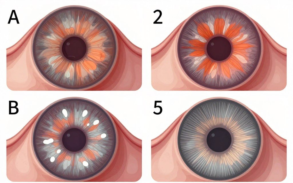
Unusual colors appearing in the kidney zone require careful interpretation. Orange or rust-colored deposits may indicate kidney stone formation tendencies. Dark brown or black spots could suggest toxic accumulation.
White or very light areas sometimes appear in kidney zones. These represent acute inflammatory responses or irritation. Yellow-green tinges can indicate liver-kidney interaction problems.
| Sign Type | Appearance | Possible Indication | Clinical Correlation |
| Orange Pigment | Rust-colored deposits | Mineral imbalance, stone formation risk | Calcium oxalate tendency |
| Dark Brown Spots | Concentrated melanin areas | Toxic accumulation, heavy metal exposure | Reduced filtration efficiency |
| White Markings | Bright, reflective areas | Acute inflammation, infection response | Active kidney irritation |
| Deep Lacunae | Dark holes in tissue | Constitutional weakness, genetic predisposition | Chronic kidney vulnerability |
| Radial Furrows | Lines extending through zone | Nerve supply deficiency, energy depletion | Functional kidney weakness |
Topographical Changes
Some kidney zones show altered topography compared to surrounding iris areas. Raised tissue elevation suggests chronic congestion or inflammation. Depressed areas indicate tissue atrophy or functional decline.
Fiber direction changes also provide diagnostic information. Twisted or spiraled fibers in the kidney zone suggest distorted energy flow. Broken or separated fibers indicate structural weakness requiring attention.
Professional Interpretation Techniques for Iridology Chart Left Eye
Accurate kidney marker assessment requires systematic examination procedures. Professional iridologists follow specific protocols to ensure reliable results. These techniques minimize interpretation errors and increase diagnostic accuracy.
Systematic Examination Protocol
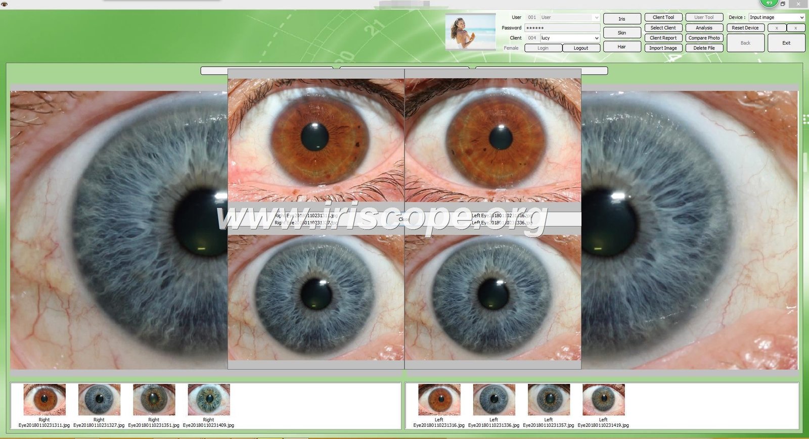
Begin examination under proper lighting conditions. Natural daylight or full-spectrum lighting works best for revealing true iris colors and structural details. Magnification helps identify subtle changes that naked eye examination might miss.
Professional-grade iridology cameras capture high-resolution images for detailed analysis. Advanced systems, including those with Mac OS-compatible software, enable precise documentation and comparison over time. Quality imaging equipment proves essential for professional practice. Learn more about professional iridology camera systems designed for clinical applications.
- Position patient comfortably with head stabilized
- Ensure adequate illumination without causing pupil constriction
- Capture multiple images at different focus depths
- Examine both eyes for comparative assessment
- Document findings with precise location notation
- Compare current images with previous examinations when available
Contextual Assessment Approach
Never interpret kidney markers in isolation. Consider the entire iris chart context and surrounding zones. Related areas include adrenal glands, bladder, and urinary tract positions.
Correlate iris findings with patient history and symptoms. The left eye iridology map provides clues, not definitive diagnoses. Professional practitioners integrate iris analysis with comprehensive health assessments.
Important Consideration: Iridology serves as a complementary assessment tool. It does not replace conventional medical testing for kidney function. Always recommend appropriate laboratory tests when kidney problems appear indicated.
Documentation Best Practice
Maintain detailed records of all iris examinations. Photograph both eyes during each session. Store images with proper patient identification and examination date.
Create standardized notation systems for describing findings. Consistent terminology ensures clear communication with other practitioners. Digital record systems facilitate long-term tracking of changes iris patterns over months and years.
Clinical Correlations and Health Problems
Specific iris markers correlate with recognized kidney conditions. Understanding these connections helps practitioners provide relevant health guidance. Research continues exploring relationships between iris signs and clinical pathology.
Common Kidney Conditions Reflected in Iris
Chronic kidney inflammation often shows as white or yellowish discoloration in the kidney zone. The brightness indicates active irritation and immune system response. These signs may precede detectable laboratory abnormalities.
Kidney stone formation tendency appears through specific pigmentation patterns. Orange-brown deposits in the kidney area suggest mineral imbalance and crystallization risk. Some iridologists report correlation between these signs and confirmed stone history.
Systemic Conditions Affecting Kidneys
High blood pressure impacts kidney health significantly. Iris signs of hypertension include vascular changes visible throughout the iris. The combination of cardiovascular markers plus kidney zone abnormalities suggests blood pressure effects on renal function.
Diabetes affects kidney filtration over time. The pancreas zone appears at 7 o’clock on the left iris chart. When both pancreas and kidney zones show abnormal signs, diabetic kidney complications warrant consideration.
Acute Kidney Issues
Sudden kidney problems produce distinct iris changes. Bright white markings indicate acute inflammation. Sharp, well-defined lesions suggest recent onset conditions.
- Sudden appearance of white inflammatory markers
- Sharp contrast between affected and surrounding tissue
- Possible pupillary response changes
- Associated adrenal zone reactivity
Chronic Kidney Degeneration
Long-standing kidney decline shows through progressive structural deterioration. Deep lacunae indicate constitutional weakness. Dark pigmentation suggests toxic burden accumulation.
- Gradual fiber structure breakdown over time
- Increasing tissue discoloration and density changes
- Progressive lesion development and deepening
- Spreading of abnormal patterns to adjacent zones
Limitations of Iris-Based Assessment
Iris examination cannot diagnose specific disease states with certainty. It reveals tendencies, weaknesses, and constitutional factors. Clinical testing remains necessary for confirming kidney dysfunction.
No research demonstrates that iridology can detect early-stage kidney disease more effectively than laboratory tests. Standard medical evaluation should always complement iris analysis. Professional iridologists recognize these boundaries and practice accordingly.

Historical Development of Kidney Mapping in Iridology
The placement of kidney zones on iris charts evolved through decades of clinical observation. Early iridology pioneers established basic organ positions. Later practitioners refined these maps through systematic patient study.
Contributions of Ignaz von Peczely
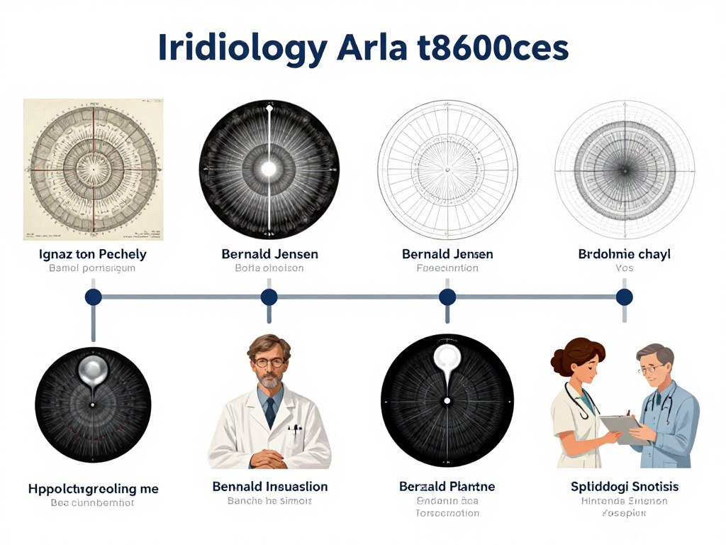
Ignaz von Peczely created the first comprehensive iris chart in the 1860s. This Hungarian physician observed iris changes in an owl with a broken leg. He theorized that the iris reflects conditions throughout the body.
Von Peczely conducted extensive patient observations to map organ locations. His original chart placed kidneys in positions similar to modern maps. Subsequent research validated and refined his initial observations.
The work of Ignaz von Peczely laid foundations for contemporary iridology practice. His systematic approach established iridology as a structured diagnostic system. Modern practitioners still reference his pioneering contributions.
Evolution Through Bernard Jensen
Bernard Jensen advanced iridology significantly during the twentieth century. This American naturopath studied thousands of patients over fifty years. His detailed observations produced refined iris mapping systems.
Bernard Jensen confirmed the 5 o’clock position for the right kidney on left iris charts. He documented specific signs associated with various kidney conditions. His published works remain standard references for modern iridologists.
Jensen emphasized constitutional assessment through iris analysis. He taught that iris patterns reveal inherent strengths and weaknesses in organs. This approach shaped how contemporary practitioners interpret kidney zone markings. Explore comprehensive Bernard Jensen iridology principles and their modern applications.
Modern Research and Validation Efforts
Contemporary research examines iridology claims through scientific methodology. Some studies show correlations between iris signs and health conditions. Other research questions the diagnostic reliability of iris analysis.
The stability iris patterns throughout life supports constitutional assessment applications. Genetic factors influence basic iris structure. Changes over time may reflect acquired health conditions versus inherited predispositions.
Biometric technology now enables precise iris pattern analysis. Advanced imaging systems document minute details invisible to earlier observers. This technology may reveal new correlations between iris features and systemic health.
Practical Applications in Professional Practice
Professional practitioners integrate left iris kidney assessment into comprehensive health evaluations. This information guides wellness recommendations and lifestyle modifications. Practical application requires skill development through training and experience.
Client Education Strategies
Explaining kidney zone findings to clients requires clear, understandable language. Avoid creating unnecessary alarm about minor iris variations. Present findings as information for health awareness rather than definitive diagnoses.
Visual aids help clients understand their iris patterns. Showing them their own iris images alongside standard iridology chart left eye diagrams clarifies explanations. People often find seeing their own eyes fascinating and educational.
Effective Communication Approaches
- Use clear, jargon-free terminology
- Provide visual comparisons with standard charts
- Explain findings in context of overall health picture
- Offer specific, actionable wellness recommendations
- Emphasize preventive health opportunities
- Encourage appropriate medical testing when indicated
Communication Pitfalls to Avoid
- Making definitive disease diagnoses from iris alone
- Creating excessive worry about minor variations
- Overstepping professional scope of practice
- Ignoring need for conventional medical evaluation
- Making exaggerated claims about iridology capabilities
- Dismissing client concerns or symptoms
Integration with Other Assessment Methods
Combine iris analysis with comprehensive health history taking. Ask specific questions about kidney function symptoms. Inquire about urination patterns, fluid retention, and lower back discomfort.
Recommend appropriate laboratory testing when iris findings suggest kidney concerns. Standard kidney function tests include serum creatinine, blood urea nitrogen, and urinalysis. These objective measures complement iris assessment information.
Consider nutritional assessment in relation to kidney health. Evaluate dietary protein intake, mineral balance, and hydration habits. The left eye iridology chart provides context for targeted nutritional recommendations.
Wellness Planning Based on Kidney Zone Findings
Develop individualized wellness plans addressing identified kidney zone concerns. Recommendations might include increased water intake, reduced sodium consumption, or specific herbal support. Tailor suggestions to each person’s unique situation.
Monitor progress through follow-up iris examinations. Document any changes iris patterns over time. Improvements in kidney zone appearance iris may correlate with enhanced overall health and successful lifestyle modifications.

Frequently Asked Questions About Kidney Markers on Left Iris Chart
Why does the right kidney appear on the left iris instead of the right iris?
The iris maps follow a cross-over principle where each eye reflects the opposite side of the body. The left iris shows right-side body organs, while the right iris displays left-side structures. This pattern relates to neural pathway crossover in the brain and nervous system connections. All organ positions on iridology charts follow this consistent cross-lateral arrangement established through decades of clinical observation.
Can iridology detect kidney disease before medical tests show abnormalities?
No scientific evidence demonstrates that iridology can diagnose kidney disease earlier than standard medical testing. Iridology may reveal constitutional weaknesses or tendencies toward kidney problems. However, it cannot replace laboratory tests like serum creatinine, GFR calculations, or urinalysis for detecting active kidney dysfunction. Professional iridologists use iris assessment as one tool among many in comprehensive health evaluation, always recommending appropriate medical testing when kidney concerns arise.
What training do practitioners need to accurately interpret kidney markers on iridology map left eye?
Competent kidney zone interpretation requires comprehensive iridology training covering anatomy, iris structures, and systematic assessment protocols. Quality programs include hundreds of hours studying iris patterns, practicing with supervised case study work, and learning from experienced instructors. Many practitioners pursue iridology certification through established professional organizations. Continuing education helps practitioners stay current with evolving research and interpretation methods. Practical experience examining numerous iris examples builds skill over time.
Do kidney markers change over time as health improves or declines?
Basic iris structure remains relatively stable throughout life. Constitutional markers like fiber patterns and major lesions generally persist unchanged. However, some iris features do shift with health changes. Pigmentation can lighten or darken. Acute inflammatory signs may appear during kidney stress and fade with resolution. The stability iris structure provides means for tracking chronic versus temporary conditions. Regular photographic documentation enables objective comparison of changes iris appearance over months and years.
Are there any side effects or risks from iris examination?
Iris examination itself presents no physical risks or side effects. The process simply involves looking at the eye under magnification and proper lighting. No contact with the eye occurs during examination. Photography uses standard light levels that don’t harm vision. The main consideration involves proper professional boundaries – practitioners should not make claims beyond their training or discourage appropriate medical care. Iris analysis serves as an informational tool, not a replacement for conventional medical diagnosis and treatment.
Can medications or supplements change the appearance iris kidney zone?
Some substances may affect iris pigmentation over extended use. Long-term medication use occasionally produces drug deposits visible in iris tissue. However, these changes typically appear gradually over years rather than weeks or months. Most nutritional supplements don’t create visible iris modifications. Acute kidney support interventions rarely produce detectable iris changes. Constitutional fiber patterns and structural features remain unaffected by medications or supplements. Focus iris assessment on stable, long-term markers rather than expecting rapid visible changes from interventions.
Professional Equipment for Iridology Chart Analysis
Accurate kidney marker assessment depends on quality examination equipment. Professional-grade tools reveal details impossible to see with simple magnification. Investment in proper equipment elevates practice standards and diagnostic capability.
Essential Equipment Components
High-resolution digital iridology cameras form the foundation of modern iris assessment. These specialized devices capture iris images at magnifications revealing fiber structure and subtle color variations. Quality cameras include proper illumination systems that don’t cause excessive pupil constriction.
Dedicated iridology analysis software enhances examination accuracy. Professional programs display captured images alongside standard iris chart overlays. Advanced systems support both Windows and Mac OS platforms, providing flexibility for different practice environments.
Camera System Features
Modern systems offer multiple advantages over basic examination methods. High-resolution sensors capture microscopic details. Adjustable lighting ensures optimal tissue visualization.
- Minimum 5-megapixel sensor resolution
- Adjustable LED illumination systems
- Focus control for different iris depths
- USB connectivity for computer integration
- Comfortable patient positioning options
Software Capabilities
Analysis software transforms raw images into actionable information. Professional programs include comprehensive chart libraries and documentation tools.
- Digital chart overlay functionality
- Patient record management systems
- Before-after comparison tools
- Report generation capabilities
- Multi-language support options
Chart Reference Materials
Comprehensive chart collections support accurate interpretation. Multiple mapping systems accommodate different methodological approaches.
- Standard left iris chart diagrams
- Right eye comparative charts
- Organ zone detail illustrations
- Pathological marker reference guides
- Digital and printed format options
Advantages of Advanced Technology
Professional equipment enables documentation of patient progress over time. Stored images provide objective records of initial findings and subsequent changes. This capability strengthens professional credibility and supports evidence-based wellness planning.
Quality imaging systems also facilitate collaboration among practitioners. Shared images enable consultations with colleagues for complex cases. Educational applications benefit from high-quality examples for training new iridologists.

Professional Partnership Opportunities for US Market
MAIKONG welcomes qualified practitioners, wellness centers, and health industry professionals as authorized distributors and agents throughout the United States. With over twenty years of manufacturing expertise in professional iridology equipment, we offer factory-direct advantages that support your practice growth and client satisfaction.
Our comprehensive product line includes advanced iriscope systems, high-resolution iridology cameras, professional analysis software compatible with both Windows and Mac OS platforms, and complete iridology chart collections. We provide multi-language software support, OEM customization options, and comprehensive technical training.
Partner Benefits
- Factory-direct competitive pricing structures
- Comprehensive product training and certification
- Ongoing technical support in your local language
- Marketing materials and professional resources
- Exclusive territorial distribution rights available
- Custom branding and OEM opportunities
Ideal Partner Profile
- Established iridology practitioners and training centers
- Naturopathic clinics and holistic health facilities
- Medical equipment distributors and importers
- Health industry entrepreneurs and investors
- Professional organizations and educational institutions
- Wellness product retailers with professional clientele
Advanced Interpretation Considerations
Experienced iridologists recognize that kidney zone assessment involves nuanced interpretation beyond basic marker identification. Several factors influence how practitioners evaluate findings and develop recommendations.
Constitutional Type Assessment
Basic iris color and fiber density indicate constitutional strength. Dense fiber structures suggest robust genetic foundations. Looser fiber arrangements may indicate inherent vulnerabilities requiring extra support.
Blue-eyed individuals with loose fiber structure face different health challenges than brown-eyed people with tight fibers. The kidney zone appearance must be interpreted within this constitutional context. Identical markings carry different significance in different constitutional types.
Bilateral Comparison Importance
Always compare kidney zones in both eyes. The left kidney appears at 5 o’clock on the right iris. Examining both kidneys reveals whether issues affect one kidney or both.
Asymmetrical findings between left and right kidney zones suggest unilateral problems. Bilateral similar markings indicate systemic influences affecting both kidneys. This distinction guides more targeted wellness recommendations.
Understanding right eye iridology chart mapping complements left eye analysis for comprehensive bilateral assessment.
Relationship to Stress Patterns
Kidney function responds significantly to chronic stress. Stress rings in eyes, also called nerve rings, appear as concentric circular lines in the peripheral iris. Their presence indicates prolonged stress impact on body systems.
When stress rings appear alongside kidney zone abnormalities, stress management becomes a priority recommendation. The combination suggests that stress contributes to kidney strain. Addressing stress may improve kidney zone appearance over time.
Learn more about identifying and interpreting stress rings in eyes as part of comprehensive iris assessment.

Conclusion
The right kidney position at 5 o’clock on the left iris chart provides valuable information for professional iridologists. Accurate identification and interpretation of kidney zone markers requires proper training, quality equipment, and systematic assessment protocols.
Understanding normal versus pathological signs in this critical zone supports comprehensive health evaluation. Professional practitioners integrate kidney zone findings with overall iris assessment, patient history, and appropriate medical testing.
Continued education and practice refinement enhance interpretation accuracy. The field of iridology evolves as new research emerges and technology advances. Committed practitioners stay current with developments while maintaining clear professional boundaries and ethical standards.
Quality examination equipment, including advanced imaging systems and comprehensive analysis software, elevates professional practice capabilities. Investment in proper tools demonstrates commitment to providing clients with the most accurate and detailed assessments possible.
Professional Support and Contact Information
MAIKONG provides comprehensive technical support for professional iridologists and wellness practitioners throughout the United States and internationally. Our experienced team assists with equipment operation, software functionality, and interpretation consultation.
Technical Support
For assistance with iridology equipment, software operation, or chart interpretation questions, our technical specialists are available via WhatsApp.
WhatsApp Support:
+86-13510907401
Available for real-time consultation regarding equipment setup, image capture techniques, and analysis software features.
Business Inquiries
For partnership opportunities, distributorship information, or professional equipment purchases, contact our business development team.
Email:
Lucy@iriscope.org
Website Resources:
iriscope.org
Visit our comprehensive online resource center for product information, educational materials, and professional support documentation.
Additional Resources
- About MAIKONG – Company history and manufacturing expertise
- Understanding Iridology – Comprehensive practice overview
- Iris Diagnosis Methods – Professional diagnostic techniques
- Becoming an Iridologist – Professional development pathways
- Factory Certifications – Quality standards and manufacturing credentials
- OEM Services – Custom branding and product development
- Contact Information – Complete contact details and inquiry forms