iridology camera what it?
иридологическая камера

иридологическая камера

иридологическая камера

иридологическая камера
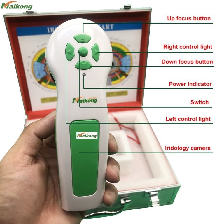
2018 newest protable USB 12.0 MP CCD iridology camera-Type:iridology camera, ProtableCertification:CEPlace of Origin:Guangdong, China (Mainland)Brand Name:QianheModel Number:QH-990UName:iridology cameraPixels:12.0 Mega pixelsColor:White and redOerate:EasyMax resolution:2560×1920OS:WIN2000, 2003, Vista, Win7,8 ,10 package Box:AluminumPackage Size:31.0 * 33.0 * 312.0cm
Общий вес: 2,0 кг
2018 newest protable USB iridology camera 12.0 MP CCD
Introduction of 12.0 MP iridology camera
Система анализа радужной оболочки глаза: международная технология, уникальные функции.
* Система анализа радужной оболочки глаза — это медицинский инструмент, который проверяет состояние тела и
предотвращает возникновение заболеваний.
* Мы привезли передовую технологию анализа радужной оболочки глаза из Германии, чтобы возглавить
люди, чтобы обнаружить источники болезней, и в любом случае заботиться о здоровье тела и духа.
* Прибор может показывать состояние тела клиентов и предлагать
клиенты получают подходящую здоровую пищу и планируют заботиться о своем теле.
1. Усовершенствованная технология анализа автоматической диафрагмы, обеспечивающая наилучшую помощь новичкам в обучении.
2) Программное обеспечение простое в использовании, поможет вам управлять.
3)Рекомендуем использовать разрешение 1024X768, оно будет лучшим.
4)Recommend to use WIN2000, 2003, Vista, Win7,8 ,10 system.
Feature of 12.0 MP CCD iridology camera
* Nice appearance and innovative design * LED illuminator around lens * Imported lens with plated layer * 12.0 Mega pixels high resolution CCD sensor * Special DSP image processor, Optical Image Stabilizer * Single capture button and digital pause capture. * Adjustable focus to give clear image. * Auto white balance and contrast adjustment, Color Temperature Filter * Dual image compare function * 3D-Negative capture mode * Compatible with iris lens, hair lens. * Deliver clear and accurate images. * Easy to operate. * Maximum resolution: 2560×1920
* OS: , WIN2000, 2003, Vista, Win7,8 ,10
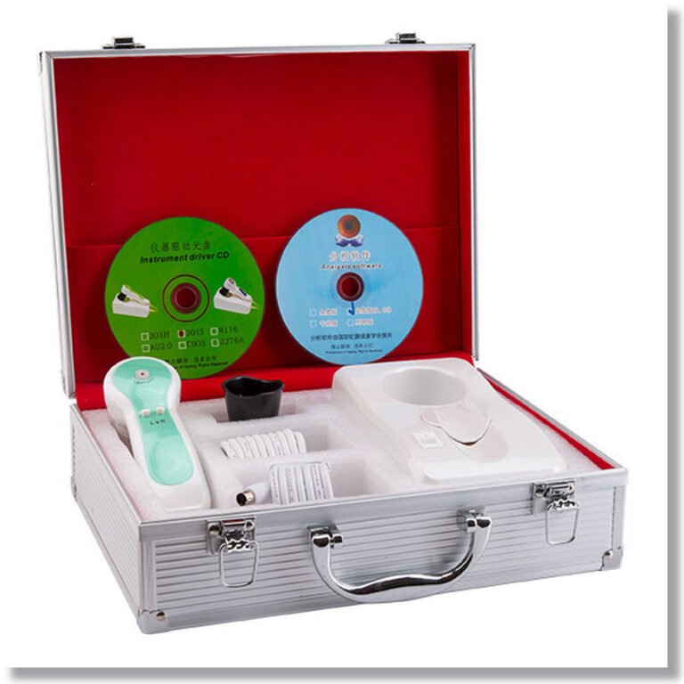
Accessories of 12.0 MP iridology camera
иридологическая камера
Рука
1 шт.
30-кратная ирисовая линза
1 шт.
Алюминиевая коробка
1 шт.
USB-линия длиной 1,5 метра
1 шт.
Защитное покрытие объектива
1 шт.
ИРИДОЛОГИЧЕСКАЯ КАРТА
1 шт.
Инструкции & Гарантия
1 шт.
Компакт-диск (драйвер и программное обеспечение Pro Analysis)
1 шт.
Want more information about the iridology camera, pls contact us:

иридологическая камера

иридологическая камера

иридологическая камера

иридологическая камера
iridology camera how to take the images?
How To Take Your Iris Photos by iridology camera-To take the best photos for your reading, set your camera to MACRO and try, if possible, to use natural, daytime indoor light with a flash. Set the size of the photo for a higher resolution, with a minimum of 1.2M (2208 x 1248). 1.2M (2784 x 1568) is best.
Шаг 1: Сделайте свои фотографии радужной оболочки с помощью цифровой камеры
Установите камеру на настройку макроса.
Increase Resolution to 1.2M (2784 x 1568).
Включите вспышку.
Используйте крытый дневной свет.
Встаньте в сторону из любого окна (обращение к окну вызовет блики).
Попросите кого -нибудь еще удерживать камеру или использовать штатив и таймер.
Держите верхние и нижние веки открытыми, чтобы сделать всю радужную оболочку видимой.
Снимайте одну фотографию каждой радужной оболочки за раз.
Держите глаз рядом с камерой. На макросложении вы можете находиться в 4-5 дюймах от объектива.
Шаг 2: Проверка ваших фотографий на предмет освещения и ясности
Проверьте фото на видоискателе вашей камеры. Используйте функцию Zoom, чтобы увидеть радужную оболочку.
Убедитесь, что радужная оболочка ясна; В противном случае попробуйте еще раз.
Убедитесь, что нет красного глаза; В противном случае включите «снижение красных глаз» и попробуйте еще раз.
Убедитесь, что вся радужная оболочка видна; В противном случае попробуйте еще раз.
Убедитесь, что на радужной оболочке нет существенного взгляда; В противном случае поверните свое тело немного от любого окна и попробуйте еще раз.

Отчеты об анализе иридологии

Отчеты об анализе иридологии


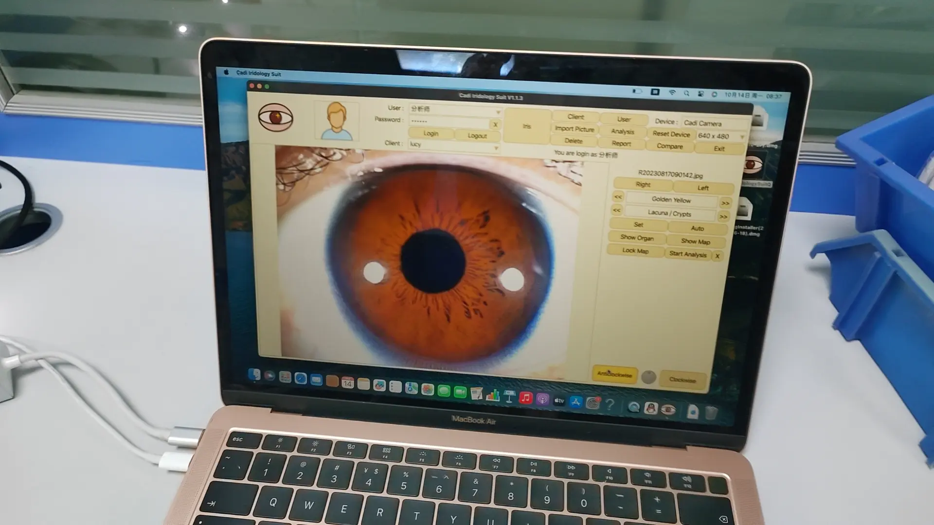
программное обеспечение для иридологической камеры

программное обеспечение для иридологической камеры

программное обеспечение для иридологической камеры

Шаг 3: По электронной почте ваши последние результаты фотографий Iris
Вы можете обрезать фотографии, чтобы вид только глаза, чтобы уменьшить размер файла.
Если это слишком много работы, то просто напишите всю фотографию.
Вы можете отправить 3-5 фотографий левого глаза в одном электронном письме.
Вы можете отправить 3-5 фотографий правого глаза в другом электронном письме.
Использование цифровой камеры: видео инструкции о том, как сделать фотографии радужной оболочки
Using an iPhone: Video Instructions on How to Take Iris Photos
Examples of Unacceptable Iris Photo Submissions
NO! Both of these example have significant glare, making portions of the iris unreadable
In the first example above, the person was likely facing a window, causing the glare to appear in the iris. The solution: Turn slightly away from the window and try again.
In the second example, it is likely that this photo was taken at night or in a room with no windows and only overhead light. Due to the darker light in the room, the light is refracting off the iris, causing significant glare and making the photo blurry. The solution: Take the photo in indoor daylight with no overhead lighting. Side lighting is usually ok.
A small amount of glare in the pupil (the black dot in the center of the iris) is ok.
NO! Not looking directly at the camera lens creates a distorted image of the iris
In the above 2 examples, the individuals are most likely trying to take the photos themselves so they are inadvertently looking at the camera while trying to take the photo.
The solution: Have someone else hold the camera steady for you or use a tripod with a timer.
NO! In these photos, the top and/or bottom of the iris is covered
When taking your photographs, check to see that the entire color portion of the eye is visible, especially the top and bottom. If you tend to have ‘droopy’ eyes, just gently pull the skin away from the eye using your thumb and forefinger.
Examples of Acceptable Iris Photo Submissions
YES!! Perfect photos – note flash inside pupil and full iris visible
YES!! Although not perfectly clear photos, these 2 examples are still readable
YES!! Very good photos – full iris visible, clear and easy to read for Iridology
YES!! Perfect photos – note flash inside pupil and full iris visible
YES!! Perfect photos – note flash inside pupil and full iris visible
Learn what to expect from your Iridology Analysis at Iridology Explained.
Find answers to any additional questions about Iridology and how it works at Iridology – FAQ’s.
Start Now! Schedule your online appointment at Book Your Iridology Consultation.
Iridology cannot diagnose specific disease.


iridology camera software what can do?
1) Добавить информацию Comstomer в программное обеспечение.
2) Редактировать или удалить информацию Comstomer.
3) Возьмите изображения глаз иридологии в программное обеспечение.
4) Анализ изображения иридологии глаз.
5) Пинта Иридологический анализ отчетов.
6) Сравнение до и после иридологических изображений глаз.
7) Сравнение отчетов по анализу иридологии.
Как использовать HE CADI CV Advance Analysess System English Version Auto Iridology Software?
1) Откройте на рабочем столе “Система расширенного анализа CadiCV, английская версия”
2) Используйте Select “пользователь”, пароль: 111111 и нажмите: “авторизоваться”
3) Нажмите “Клиентский инструмент”, введите информацию о вашем клиенте. Если хорошо, нажмите “добавлять”,и
Клик”закрывать”
4) Нажмите “захватить правый глаз”.–щелкнуть “захватывать”, Повторение левого глаза последним шагом.
5) Выберите картинку глаз (рис.
6) Нажмите “анализ”
7) Нажмите “Установите параметр” кнопка.
1 -й Поместите стрелки мыши в центр зрачка, нажмите «Левый ключ из мыши»,
2 -й удалить стрелки края мыши ученика, нажмите «Левый ключ из мыши»,
3rd. Сделайте стрелки мыши между радужной оболочкой и склерой, затем нажмите «Левый ключ из мыши».
И нажмите кнопку «Установить параметр».
8) Нажмите “Анализ радужной оболочки” Кнопка и нажмите кнопку «Показать орган».
Затем выберите, какую часть глаз вы хотите анализировать, просмотреть его цвет и
Форма, наконец, нажмите на его цвет и форму, как следующий «коричневый, комок»,
Удалите стрелки мыши в вашу правильную часть, наконец, вы можете увидеть следующее
изображение результата анализа, если вы выбираете «цвет или форма», неверно, нет никакого результата детали
ПРИМЕЧАНИЕ. Когда вы можете прочитать результат анализа каждый раз, нажмите «Добавить в отчет» правого глаза, магазина
результат анализа. Конечно, вы можете продолжить любую часть правого глаза, повторить, чтобы выбрать цвет и
форма.
Когда вы закончите анализ правого глаза, пожалуйста, нажмите «Анализ», затем выберите «Левый» глаз, чтобы
Повторяйте выше операции.
9). Когда вы закончите анализ всех деталей, нажмите «Анализ» еще раз, затем нажмите «Сохранить», чтобы сохранить
Полный результат анализа радужной оболочки клиентов.
10) Кнопка нажмите кнопку «Отчет о клиенте» и нажмите свое имя,

cadi iridology software

cadi iridology software
iridology camera manaul
IR-9915U iridology camera for sale


