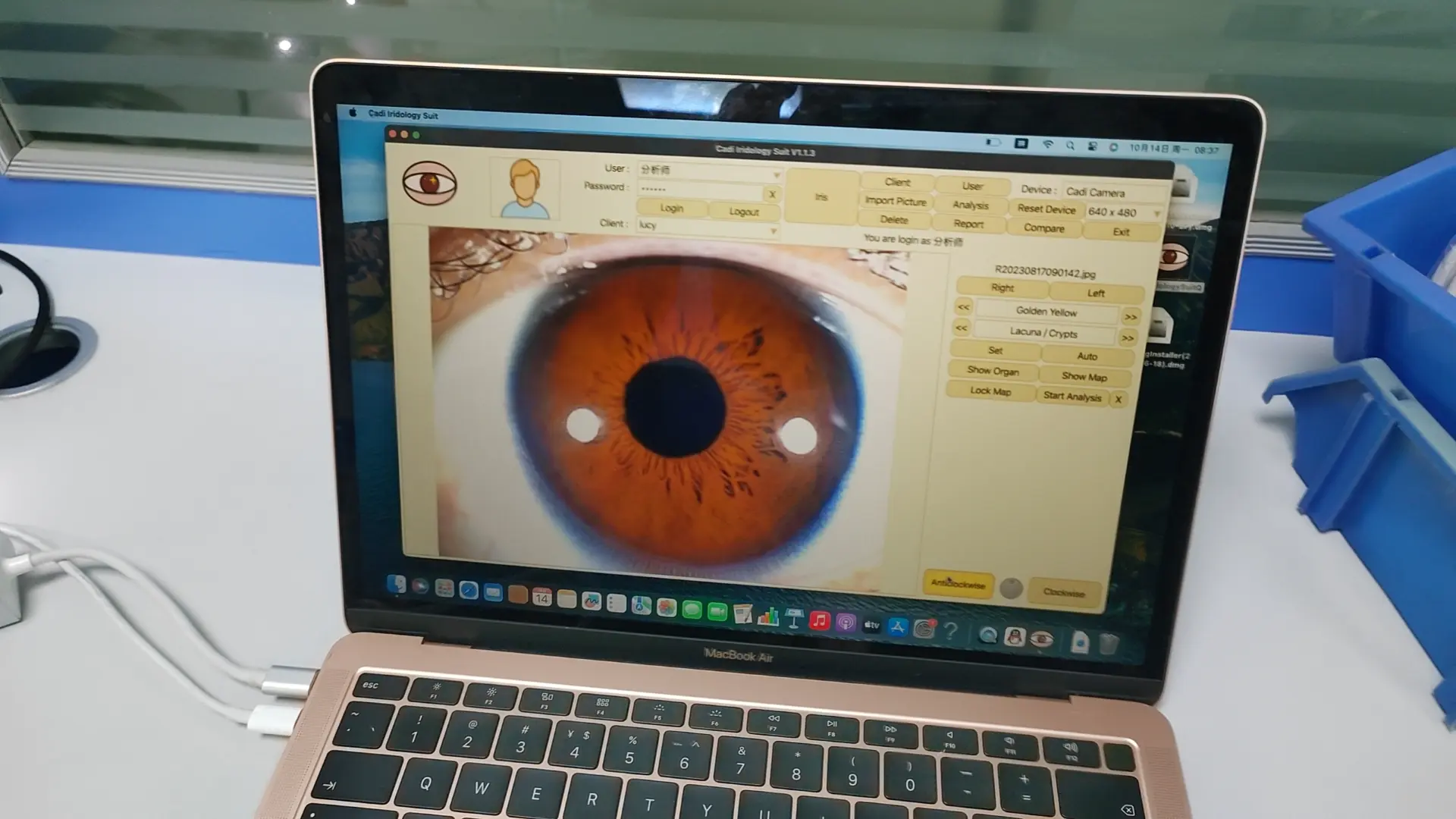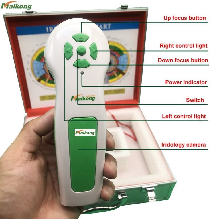
SD8004 Super Digital iridology camera for mac
SD8004 Super Digital iridology camera for mac
Joyful Living Services, in partnership with Iris Imaging, is excited to announce that we now offer an 24.2MP Digital Iridology and Sclerology Camera System that has the capability of extremely high resolution and uncompromising quality at affordable prices.
Our Iridology and Sclerology System is based on the 24.2MP Nikon DSLR. The system uses a highly rated, crystal clear 90mm lens that has even greater clarity than the 105mm macro lens for unparalleled performance and clarity.
The camera features a newly designed oversized Nikon CMOS sensor specifically optimized for low noise and high saturation, giving you the best image quality available. It also uses Image-processing engine EXPEED 3 – the same as that used by the Nikon D4 FX-format flagship camera and a 3-inch Clear View LCD with anti-reflective and scratch-resistant coating. You’ll have uncompromised Digital performance with power and flexibility in the palm of your hands.
We use a highly regarded macro lens along with a specifically designed flash and two high quality fiber optic strands to illuminate the eye perfectly. This allows for greater depth of field and consistently clearer pictures. The flashes are still within the pupil so no part of the eye is obscured.
The portable fully adjustable custom camera stand is made of heavy duty plate aluminum with a beautiful “Brite Dipped” face mount rather than the plastic found in other systems. This gives you unequaled beauty, strength, stability and longevity. It is easy to set up on any table, at any location for ultimate portability and comfort.
The lighting system gives you perfectly illuminated shots every time without flash spots on the iris. The twin head focus light is embedded in the flash ring giving you ease in focusing while being easy on the client.

What is 12MP iridology camera for mac?
What is 12MP iridology camera for mac?
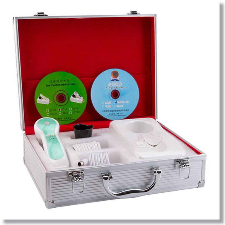
Accessories:
1. Handset X1pc
2. 30X Iris Lens X1pc
3. Leather PU Box X1pc
5. 1.5 Meter USB Line X1pc
6. Lens protective cover X1pc
Package:
* IRIDOLOGY CHART X1pc
* Instructions & Warranty X1pc
* CD (Driver and Pro Analysis Software) X1pc
What is the primary benefit of photo iridology ?
Not only does it bring clues to your iridology analysis, but it helps to create a better connection with your patient. The patient, who’s naturally curious, is eager to see their own iris. With some simple explanations they can get a basic understanding of your analysis. The connection with your patient strengthens and their understanding of their process improves.
* Iris analysis system: international technology, unique functions.
* Iris analysis system is a medicinal tool that checks the body conditions and prevents diseases from occurring.
* We brought in the advanced iris analysis technology from Germany to lead people to discover sources of illness, and care the body health and spirit in anyways.
* The instrument can show the body conditions of customers and suggest customers the suitable health food, and the plans to care their bodies.
Healing With Iridology:
The perfect iris has never been seen and all individuals exhibit some degree of tissue weakness, whether it was acquired and developed from questionable health habits and environment, or from parents genetic makeup.
Healing comes from the root word meaning whole. So does health and holy!
Healing comes from looking after all areas of our whole self, being mind, body and spirit.
In my opinion, when you get rid of what the body can’t use and give it what it needs, the body will do the rest. The body is a wonderful incredible specimen. Everyday it breathes by itself, the heart pumps by itself, if you cut yourself or break a bone, they heal by themselves. You never have to tell your body what to do automatically. Our responsibility is to provide the elements for the body.And you are the only one who can do that.
12MP iridology camera for mac Specifications
1.High Resolution, Real 2 magepixel pictures
2. Easy operating without driver installation
3. luxury leather package
12MP iridology camera for mac Instruction:
* Nice appearance and innovative design
* LED illuminator around lens
* Imported lens with plated layer
* 8.1 Mega pixels high resolution CCD sensor
* Special DSP image processor, Optical Image Stabilizer
* Single capture button and digital pause capture.
* Adjustable focus to give clear image.
* Auto white balance and contrast adjustment, Color Temperature Filter
* Dual image compare function
* 3D-Negative capture mode
* Compatible with iris lens, hair lens.
* Deliver clear and accurate images.
* Easy to operate.

Confessions of a Former iridology camera for mac
Confessions of a Former iridology camera for mac
I am a former iridologist. I did not abandon iridology and much of “alternative medicine” lightly. It was a decision that I struggled with, but my conscience forced me to make the fateful choice. When I abandoned the field I lost my income and my identity. It was a difficult choice, to say the least. How did I get involved with “alternative medicine”, and what prompted my decision to forsake a field that was such an important part of my life?
Let’s Start At the Very Beginning
I was born into a family that had a natural interest in “alternative medicine”. When I was around the age of seven my parents stumbled onto the practice of iridology in their quest to find a cure for my mother’s cancer. The practice fascinated me and as my father began to research iridology I learned all I could from him. I eventually received my own certificate from an iridology-training program. As early as age nine I had read Jim Jenks’ book “The Eyes Have It” and was attempting to tackle Dr. Bernard Jensen’s various works on iridology. I learned to look into the iris, the colored part of the eye, and determine an individual’s health needs.
The theory is that the iris contains nerve fibers connected to various parts of the body through a previously unknown nerve pathway in the cranial nerves. Ignatz Von Peczely, a 19th century Hungarian, reportedly began the study of iridology after noticing changes in patients’ irises. Proponents claim:
The information from each organ of the body is relayed to the iris via the oculomotor nerve (cranial nerve III).
The health value of each organ and organ area is determined by examining the color, lightness/darkness, shape, and depth of the fibers of the iris.
Generally the lighter color of the fibers, the more activity, and possibly, sensation (such pain) there is supposed to be at the tissue level of that organ.
Generally the darker an area or fiber, the less the activity in that area. For example, a bright white fiber in the lower back area would likely represent back pain currently being experienced by the owner of the eye, whereas a black area in the lower back would suggest severe back injury that has disrupted the nerve activity from that area.
In my practice I subscribed to the Bernard Jensen methods of iridology and found them quite successful. I enjoyed having skeptical clients come into my office and become believers as I told them things about themselves that they didn’t think anyone could know. I constantly researched methods to make my practice more precise and read every book I could find written by Bernard Jensen on iridology.
As is usually the case, my practice in alternative medicine was not limited to simply iridology. I was certified in three forms of applied kinesiology, was taught how to prescribe herbal therapies, used homeopathy, suggested diet changes, and engaged in emotional/spiritual counseling. I also took a strong stand against drug prescriptions, most vaccinations, and most elective surgeries. I firmly believed that medical doctors were trained in how to destroy the body while I had learned methods that really had benefit by building the body from the inside. I personally felt, and taught, that the only benefit of conventional medicine was found in severe emergencies, such as trauma.
I believed that the human body was some sort of remarkable collection of intelligent organs and systems that worked together almost magically to create a healthy unit. I believed that illness was the result of poor communication and imbalance between organ systems. I thought that herbal treatments fed the organ systems so that they could work out their kinks and create health from disease. It was my understanding that pharmaceutical treatments only tweaked the organs within a system to cover up the symptoms of a disease. For example I felt that treating a fever from a viral infection with Tylenol only covered up the infection and allowed the virus to return when the host was too weak to protect himself in the future. I felt that the best way to treat a fever was to let it run its course. I thought that the body would best treat itself if we fed it right and that herbs only provided the necessary compounds to treat the body so that it could work its “magic”.
The Beginning of a Change
I was a staunch supporter of alternative health care when I decided that a balanced approach to health care should include an understanding of conventional medicine. To that end I decided on a program provided by the Medical Training Institute of America that trained health care consultants. The basic idea of the program appealed to me in that it involved studying directly under practicing physicians in a clinical atmosphere. My hope was that the knowledge I could gain would allow me to better integrate conventional and alternative medicine.
One of the requirements for entry in the program was interview by a panel of five doctors. When these men learned of my past training in alternative medicine they balked. After some negotiation we came to an agreement and I breathed a sigh of relief as they agreed to let me in the program.
I expected more resistance as I began studying with the doctors assigned to my training. To my surprise many of the physicians were interested in alternative medicine. Once they learned of my past experience and that of my father they asked quite a few questions on various treatments we used. They would describe a sticky point in their treatment of a specific condition and then ask if we had good results with our treatment. When I answered in the affirmative they usually responded with increased interest that would deflate as I explained my answer.
I soon learned that alternative and conventional medicine had different levels of evidence and verification of treatment success. As I would explain a treatment that I thought was successful the doctors would ask questions like “What kind of studies supported this?” Or “Is that documented with blood tests?” I began taking notes on how I could document our results better as well as studies that we needed to look for or instigate.
In addition to working in a clinical setting, the health care consultant program included studies in advanced anatomy and physiology, pharmacology, biochemistry, microbiology, and histology. The study of these sciences matured my understanding of alternative medicine. For example, I now understood herbs as biochemical compounds that intervened in bodily functions in much the same way as pharmaceuticals. I returned home from my three years of training with excitement and many new ideas on how to treat and evaluate several conditions.
Though I had performed occasional iridology exams on a few individuals prior to the completion of the health care consultant program, my return home marked the beginning of what I had hoped would be a long and successful career as a naturopathic practitioner, and I officially joined my practice with my mentor’s successful practice.
So Many Imperfections
When I began working with iridology, I looked into the iris with a penlight and loupe, marking my findings on a photocopied version of Jensen’s eye chart. Eventually I began using a video camera and a monitor to record and examine irises. The video camera was large step in keeping track of changes in the iris and it also allowed me to examine an iris without getting into the patient’s face ourselves (reducing the risk of being coughed on and smelling too much bad breath). The use of the camera to record and review past visits refined my ability to see changes in the iris. When using the chart to record iris signs I found that my ability to detail exact proportions of iris signs from visit to visit was extremely variable, and left too much up to my fallible memory. The use of the video record allowed me to measure signs and detail any change.
It soon became apparent to me that the video camera had some major limitations as well. Before I explain the flaws, let me describe how we used the video camera to record the iris. The video camera itself was fastened to a device that had a cup for the patient’s chin to rest in and essentially remain stationary. The camera was affixed to a platform on the device that would allow the camera to be rolled in two planes- forward to back, and side to side. The camera was fitted with a macro lens that allowed an enlarged picture of the iris to fill the entire video screen. Each eye was recorded singly; the right being the first, with a penlight illuminating from the outside and the eyes focused on a stationary object. Each eye was recorded for 10-15 seconds with the penlight moving between two points to allow the iridologist to view parts of the iris that would otherwise be obscured by the glare of the penlight (the glare would appear as a bright white dot about 1cm in diameter).
I soon found that structure “changes” could be created on the video record by changing the angle of the light to the eye. Areas that I thought were dark would suddenly show healing lines when the position of the light changed. Thick white lines would change to thin gray lines when the light moved. More than once during this period an eminent iridologist would call me to his office and show me a change he had recorded in patient’s iris minutes after doing a spinal adjustment. After closely examining his recordings, it became obvious to me that his light position and the angle of the camera to the eye had varied from time to time causing the appearance of a change in the iris.
Not only did the light placement affect the appearance of structures; the slow draining of the batteries in the penlight changed the appearance of the eye color. If the iris was recorded using two-week-old batteries it would have a slightly yellow tint. If the iris was recorded with newly opened batteries, the iris colors would almost be washed out. The lighting in the room also had an effect on the recorded color due to the contrast the camera measured. The variables were so great that I began to entirely distrust any changes that I found in the iris while using the camera.
A New Opportunity
The solution came to me while reading another iridologist’s site. He described a specialized camera that has an excellent method of defining the ideal placement for the camera to photograph the eye. I passed the information on to my friend, who purchased the camera and allowed me to use it for my clients when I was working in his office. I was ecstatic with the opportunities the new camera presented. The camera used the same flash every time which meant the lighting would keep the color constant. The flash was always in the same place, reducing the possibility of changes in shadow placement. The camera used two intersecting lines projected on the target to indicate correct placement, thus guaranteeing that the iris would always be the same size in every picture. The film was highly refined so as to show every fiber well defined. It was most certainly a superior method of recording the iris, and I was set to have strong proof of iris changes.
I was thrilled with my first “documented” iris change until I set out to measure the fibers in the iris that showed change. It soon became apparent that the changes were actually due to changes in the angle of the camera to the eye. When I corrected for this, I found very few changes in the iris, and no changes in the actual structure.
The changes I found in a few irises were actually in the color. When I experimented with changes in angles, I found that the angle of light going into the eye, and the level of lighting in the room, had an effect on pupil size. Pupil size had a direct link to fiber size, and fiber size seemed, in some cases, to be related to colors that appeared in the iris. This was more obvious in someone who had more than one color present in his or her iris. I, for example, have brown, green, yellow, and blue appearing in my iris. In different degrees of lighting my eyes have a different appearance. It is for this reason that different people have told me that my eyes are entirely brown, green, or blue.
In a smaller way pupil size affects the appearance of color in a magnified iris. Not only does light have influence on pupil size, but the autonomic nervous system also has an influence on the pupil size, thus a person’s degree of fear or alarm can change pupil size. An iridologist purports to be capable of telling a great deal about a person based on the color of single fiber. This variable became quite important for that reason. When I corrected for all these variables, I found very few iris changes. More importantly, I found very few iris changes in people who had significant health changes in the prior months. In many of the cases where the iris seemed to have changed, it had changed inappropriate to the physical changes that had been known to occur. In other words, I discovered that the iris did not reflect the level of health in the body.
But What About All Those Incredible Findings?
I have been queried by those who have read my site as to how their iridologist was able to provide information that the patient thought no one else could know. One answer is coincidence. A small percentage of iris signs that seem obvious actually do correlate to actual health conditions. When one of these correlations is found it feeds the iridologist’s sense of success and provides a believing patient. The chance for coincidence is increased by the fact that one can have multiple signs in one organ area and that most people who seek health help have commonly occuring illnesses. Another answer is the fact that the iridologist’s best tool is general questions. As an iridology student it was drilled into me that iridology cannot diagnose disease. From the holistic (alternative medicine) view this is iridology’s strength. Alternative medicine holds that diagnosis is only a method of looking at symptoms and putting a name to them. Iridologists teach that symptoms are late signs of disease and that iridology allows them to catch imbalance in the body before it causes disease. An iridology website states that “Iridology DOES NOT DIAGNOSE DISEASE, it merely reveals tissue weaknesses, inflammation or toxicity in organs or tissues.” https://www.herbsbylisa.com/iridology.htm (Lisa Ayala, Last modified: April 22, 2001 and accessed 10/11/02)
The beauty of not having to provide a diagnosis from the eye is that the practitioner simply uses the iris to create leading questions. Suppose I had a patient who had a mark in his lung area. My first question would be “Have you ever had a problem with your lungs? Something like asthma, pneumonia, or emphysema?” If the patient could remember something like that I was considered a genius, but if there was nothing obvious I would question further. “Perhaps you have had a cold recently?” If the answer was no and there wasn’t anything obvious the next step would be to look at the bowel, which is theorized to cause lung weakness. The bowel is represented in the eye as the area directly around the pupil and is usually darker than the rest of the iris. If the bowel was dark then the obvious answer was that the patient had an unknown lung weakness resultant from the bowel. If there was no bowel problem, the last answer was that there was a genetic lung weakness that needed to be treated to prevent future problems.
Does this process prove iridology? No, in fact, it condemns iridology. If the exam brings up a past history of lung problems, it simply identifies iridology as a cumbersome method of gathering a medical history and does not prove iridology’s ability to diagnose since the iris cannot identify the nature of the problem. If the exam shows a connection to the bowel it illustrates the fact that iridology flies in the face of recognized science and medicine for the lung-bowel connection has been investigated fruitlessly in true science.
Suppose the lung weakness seen is suspected to be a genetic weakness that has not manifested. The iridologist congratulates the patient for coming in when he did. If other tests do not indicate the suspected lung weakness, the iridologist replies with the statement that iridology can pick up weaknesses before they even grow to the point that they are discernable to other methods of examination. If the patient follows the iridologist’s treatment guidelines and never develops a lung problem, the patient is congratulated for avoiding a future problem. If the patient refuses treatment for his lung problem and never develops a problem with the lung the iridologist considers it a problem that is hanging over the head of the patient. If the patient ever develops any lung problem of any kind it is attributed to the weakness found in the lung.
The question that is brought to my mind is how do we know that the iris is indicating properly a lung weakness if the sign cannot be substantiated by any other method? Furthermore, how did that sign ever come to mean lung weakness if no reliable method was able to prove it? I believe that in the final analysis, iridology is very suspect and cannot fall into the category of science. Iridology is fraught with observations that are either unsubstantiated by reliable methods or simply based on questionable anecdotal evidence. Even good scientific studies have failed in their attempts to prove the potential of iridology to pick up on signs indicating known health conditions in patients. (which should be glaring in the iris)
Is There Science in Iridology?
As my inquiry of iridology proceeded, the issue of science did come up. How could I prove to someone else that the iris was anatomically and physiologically equipped to indicate what I was taught to believe it could? Early in my iridology training I was taught that nerve impulses reach the brain from the tissues in the body and are routed to the iris through the optic nerve. When the nerve information reached the iris fibers it caused a restructuring in the iris to reflect the condition of the tissues of the body. As my training went on, my teachers came across the sad fact that the iris and optic nerve have little influence on each other. The theory then changed to state that the information reached the iris via the oculomotor nerve.
What evidence indicates that the iris can change fiber structure based on the information it receives via the oculomotor nerve? One iridologist has claimed, “We know from research using electron microscopes, that each fiber in the iris is actually a nerve sheath containing over 20,000 nerve fibers. Each of the nerve fibers travels through the central nervous system to every organ, system and area of the human body. As such, each region of the iris represents an area of the body.” (eGuide on iridology) Is there correlation for this evidence?
A histology text states, “The anterior (front) surface of the iris is irregular and rough, with grooves and ridges. It is formed by a discontinuous layer of pigment cells and fibroblasts. Beneath this layer is a poorly vascularized (blood fed) connective tissue with few fibers and many fibroblasts and melanocytes (color cells). The next area is rich in blood vessels embedded in loose connective tissue.” [Taken from page 456 of Basic Histology 8th edition by L. Carlos Junquerira, Jose Carneiro, Robert O. Kelley ISBN: 0-8385-0567-8] In other words, the iris fibers are not bundles of nerve fibers, but strands of cells that are similar to the ones in cartilage. With this “microanatomy” in mind, where is the scientific support for the idea of a structural change to the iris via the nervous system? Unless other evidence is presented, there is no support for this idea.
The whole issue of presenting an anatomic, histologic, and physiologic basis for iridology is crucially important. Up to the point that I was finally willing to question the basic ideas of iridology, my previous studies presented only a shadow of doubt. Up to this point, I was able to question the validity and value of my conclusions against iridology because other iridologists had opposite conclusions or found nothing wrong with conclusions similar to mine. It was hard to believe that something I trusted most of my life was wrong.
Support For Color Changes?
Looking back it is amazing to me that I thought iridology had any chance scientifically. Once I took a look at the basic anatomy of the eye and nervous system I knew it was impossible to make a change structurally, but what about color? Perhaps the iris did not change itself structurally, but perhaps color changes would be possible to support scientifically. I knew, as stated above, that without special equipment it would be difficult to prove or disprove meaningful color changes in the iris. The question was if anyone had done such observation and if it was found to mean anything.
In order to understand any observations on iris color change one must understand the process of developing color in the iris of the eye. The base color of the iris is made up of very dark pigmented cells that are at the underside of the iris which reflect back visual blue light, thus giving the appearance of a blue eye. In albinos, the lack of pigment allows light to reflect off the blood vessels giving a pink reflection. It is the level of pigment in the upper (exterior) levels of the iris that give variation on eye colors from blue-green to dark brown. Just as genes are influential on the level of pigmentation of the skin, so genes have influence on eye color, and the structure of the iris.
I was taught as a young iridology student that various colors in the iris were deposits of chemicals. For example, a rust spot in the iris was a small spot of an injected chemical from a vaccine, or a brilliant yellow spot was the result of a sulfur deposit in the eye from the ingestion of a sulpha drug. The fact is that these spots are collections of melanin; similar to the substance that causes freckles in the skin. Eminent iridologists have written in their works that these spots were found to contain metals and other substances in autopsies. Unless metals or other substances were injected directly in the iris this cannot be true.
Based on the fact that iridology does not reflect true anatomy, physiology, or histology of the iris, and based on the fact that iris colors are not determined by nerve input, it became ludicrous for me to believe that iris color is any indication of health in remote organs. Some iridologists suggest that eye color and fiber structure are unchanging and are useful to indicate certain predisposition to mental and physical disorders. These iridologists once again use generalities and other useless methods to describe the usefulness of their method. They unfortunately fail in their attempts at using logic and science correctly.
What is My Next Step?
Based on what I now know I can find no argument that could persuade me to return to practicing iridology. Sadly, my interactions with practicing iridologists have failed to dissuade them from practicing what I feel is a deceptive and damaging practice. Why don’t any other iridologists see the light? I am not able to answer that question accurately, but I can state that every iridologist I have gotten to know thoroughly believes that iridology is one of the best methods available for ascertaining the health of one’s body. I am sure that there are some who practice iridology simply to dupe people and make money. Plenty of income can be made with iridology, but many, if not most, iridologists charge a relatively small fee for their services. These people honestly believe that they are helping people.
Despite the pure motives of those who practice iridology, one is forced to wonder how an iridologist can practice despite all the evidence against iridology. Several reasons come into play, and the list is much longer than can be enumerated here. When I was fully persuaded of the value of iridology, I was constantly confronted with evidence against iridology. I was not dissuaded from the practice by this evidence because I had believed the illogical answers I had been given by my own teachers. To this day I cannot explain how the spell was finally broken. I do not understand fully what made the difference, but I do know that being trained in true science was a part of the equation.
I have been asked by many if my rejection of iridology translates into a rejection of all of alternative medicine. I would answer no. Iridology fits into a category of disproved alternative health care practices. These practices have no basis in real anatomy and physiology and have failed well-done trials and studies. Practices that fall into this category include applied kinesiology, reflexology, therapeutic touch, homeopathy, and the Hulda Clark cure for all diseases (zapper, etc.). There are other aspects of alternative medicine that I cannot dismiss without more comment. These may have some scientific support, but are misused or over-used in alternative medicine. Some examples in this category are herbs and supplements. I feel that herbs are actually the same as pharmaceuticals and should be treated with the same respect. I disagree with the motto “I use herbs instead” because herbs act the same way inside the body as their counterparts behind the prescription counter.
One of my biggest problems with so-called “natural medicine” is that many practitioners advise herbs without any training of any kind in the science of pharmacology. I was counted as one of these. Master herbalists suggest herbs based on historic use, personal experience, patient reports of benefits, and some sketchy information from clinical trials. Few herb studies even exist to indicate interactions in the delicate systems of the kidneys and liver. Despite these limitations herbalists, iridologists, applied kinesiologists and others advise patients on the use of multiple herbs though there is little information in their libraries as to the effectiveness, benefit, and safety of these substances.
On top of this, these poorly trained practitioners are confidently advising their clients to take herbs along with the prescription medicine suggested by their bona fide physicians. Many of these individuals advising herb use don’t know the meaning of cytochrome p-450 let alone how the system works. I am willing to bet that many would think the term to mean some new dietary supplement.
My own practice of alternative medicine became very uncomfortable to me as I learned how little I had actually been taught. I began suggesting fewer and fewer supplements, trying to keep to those that I knew were documented as safe. I eventually became so uncomfortable that I felt physically ill and drained when I completed my sessions with patients. I ultimately decided to abandon my practice and begin the process of furthering my education. That is where I am now. My goal is to be a practioner of true health care—a medical doctor. To be honest, I am sure that our conventional Western medicine is not a perfect, flawless system, but my experience in alternative medicine convinces me that conventional, evidence-based medicine is a big step up compared to the alternative.

2018 newest protable USB 5.0 MP CCD iridology camera for mac
2018 newest protable USB 5.0 MP CCD iridology camera for mac
Type:iridology camera for mac, ProtableCertification:CEPlace of Origin:Guangdong, China (Mainland)Brand Name:QianheModel Number:MK-990U
Name:iridology camera for mac
Pixels:5.0 Mega pixels
Color:White and red
Oerate:Easy
Max resolution:2560×1920
OS:WIN2000, 2003, Vista, Win7/8/10
package Box:AluminumPackage Size:31.0 * 33.0 * 35.0cm
Gross Weight:2.0kg
Newest protable USB iridology camera for mac 12.0 MP CCD
Introduction of 12.0 MP iridology camera for mac
Iris analysis system: international technology, unique functions.
* Iris analysis system is a medicinal tool that checks the body conditions and
prevents diseases from occurring.
* We brought in the advanced iris analysis technology from Germany to lead
people to discover sources of illness, and care the body health and spirit in anyways.
* The instrument can show the body conditions of customers and suggest
customers the suitable health food, and the plans to care their bodies.
1Advanced auto iris analysis technology to provide the best help for beginners to learn.
2)Software easy to use, help you to command.
3)Recommend to use of 1024X768 resolution, it will be the best.
4)Recommend to use Windows XP/2000/Vista/7 system.
Feature of 12.0 MP CCD iridology camera for mac
* Nice appearance and innovative design * LED illuminator around lens * Imported lens with plated layer * 12.0 Mega pixels high resolution CCD sensor * Special DSP image processor, Optical Image Stabilizer * Single capture button and digital pause capture. * Adjustable focus to give clear image. * Auto white balance and contrast adjustment, Color Temperature Filter * Dual image compare function * 3D-Negative capture mode * Compatible with iris lens, hair lens. * Deliver clear and accurate images. * Easy to operate. * Maximum resolution: 2560×1920* OS: , WIN2000, 2003, Vista, Win7,win8
Accessories of 12.0 MP iridology camera for mac
Handset
1pc
30X Iris Lens
1pc
Aluminum Box
1pc
1.5 Meter USB Line
1pc
Lens protective cover
1pc
IRIDOLOGY CHART
1pc
Instructions & Warranty
1pc
CD (Driver and Pro Analysis Software)
1pc
Want more information about the iridology camera for mac, pls contact us:


