Iridology charts serve as fascinating tools in alternative medicine, allowing practitioners to analyze the iris of the eye to assess potential health conditions. Unlike conventional medical diagnostics, iridology proposes that specific zones of the iris correspond to different organs and body systems. This ancient practice has evolved over centuries, with modern practitioners using detailed charts to identify patterns, colors, and markings that may indicate underlying health issues or predispositions.What Is an Iridology Chart? Origins and Structure


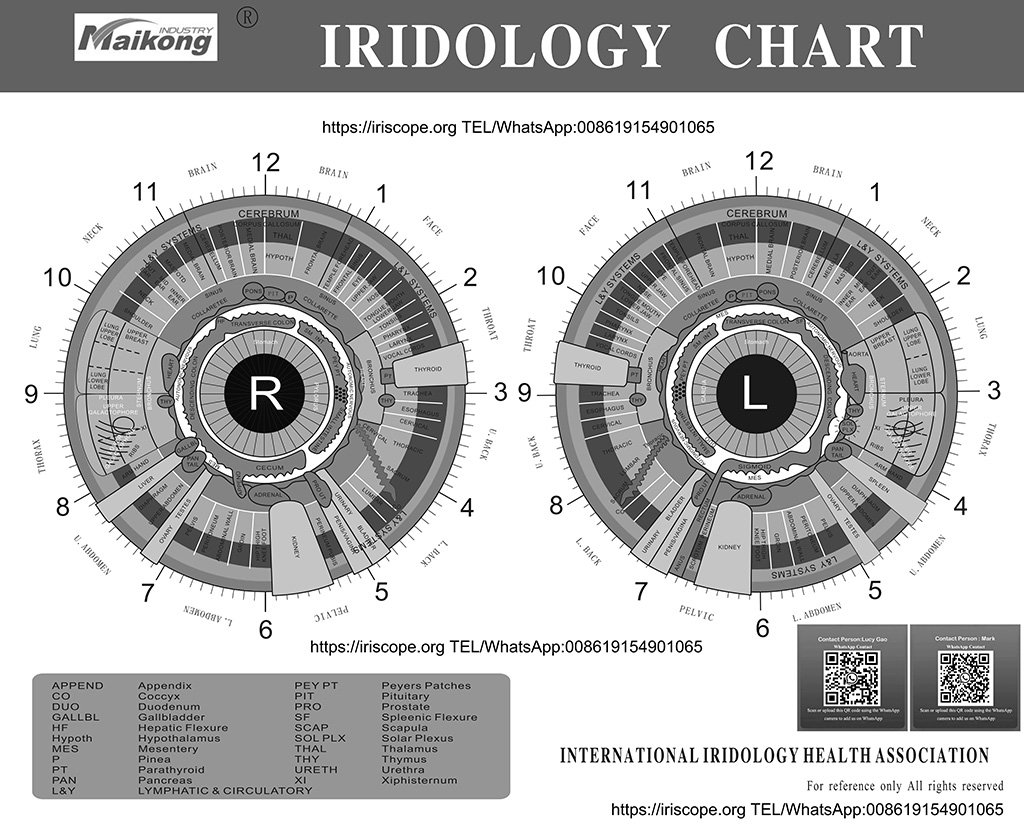
Standard iridology chart showing iris zones mapped to corresponding body systems
An iridology chart is a detailed map that divides the iris into approximately 80-90 zones, each corresponding to specific organs, glands, or body systems. This diagnostic tool emerged from the work of Hungarian physician Ignatz von Peczely in the 1800s, who reportedly noticed changes in an owl’s iris after it suffered a broken leg. The modern iridology chart has evolved significantly, incorporating detailed topographical mapping that practitioners use to analyze the eye’s structural appearance.
The fundamental structure of an iridology chart includes:
- A circular representation of the iris divided into zones like a clock face
- Radial divisions corresponding to different body systems and organs
- The right iris typically representing the right side of the body
- The left iris typically representing the left side of the body
- Concentric rings indicating different tissue layers and systems
Iridology charts serve as reference guides for practitioners to identify potential health concerns through careful examination of iris structures, colors, and markings. The practice is based on the theory that the iris connects to every organ and tissue via the nervous system, with its appearance reflecting the condition of distant body parts through neurological connections.
Download Our Free Basic Iridology Chart
Start learning about iris analysis with our beginner-friendly reference chart.
Download Free Chart
Practical Applications of Iridology Charts
Iridology charts find applications in various alternative health contexts. Here are three primary ways practitioners utilize these diagnostic tools:
1. Holistic Health Assessment

Iridologist performing a comprehensive iris examination using specialized equipment
Holistic health practitioners use iridology charts as screening tools to identify potential areas of concern before symptoms manifest. By examining the iris for specific markings such as spots, rings, or discolorations, an iridologist may detect signs of inflammation, toxin accumulation, or stress in corresponding body systems. This approach allows for preventative recommendations rather than treating existing conditions, aligning with holistic health’s emphasis on prevention and whole-body wellness.
2. Complementary Assessment in Naturopathy

Naturopathic doctor discussing iridology findings while referencing a detailed chart
Naturopathic doctors often incorporate iridology as one component of their comprehensive assessment protocols. Rather than using it as a standalone diagnostic method, they combine iris analysis with case history, physical examination, and sometimes laboratory testing. The iridology chart serves as a guide to identify potential constitutional weaknesses or genetic predispositions, helping practitioners tailor treatment plans that may include dietary modifications, herbal remedies, or lifestyle changes based on the individual’s unique patterns.
3. Educational Tool for Patient Awareness

Health educator using an iridology chart to explain body-eye connections
Beyond direct diagnosis, iridology charts serve as powerful educational tools that help patients visualize the interconnectedness of body systems. Practitioners use these visual aids to explain how lifestyle factors might affect different organs, creating a tangible connection between daily habits and health outcomes. This application transforms the abstract concept of “whole-body health” into a concrete visual representation, potentially increasing patient engagement and compliance with recommended health protocols.
These applications highlight how iridology charts function within complementary and alternative medicine frameworks. While not typically used in conventional medical settings, they remain valuable tools for practitioners who approach health from holistic perspectives that consider physical, emotional, and environmental factors.
Traditional vs. Modern Iridology Charts


Evolution of iridology charts: traditional hand-drawn (left) vs. modern digital (right)
Advantages of Modern Iridology Charts
- Higher precision with 80-90 distinct zones identified
- Digital imaging allows for tracking subtle changes over time
- Integration with software for more consistent analysis
- Color-coding systems for easier interpretation
- Standardized terminology across practitioners
Limitations of Traditional Iridology Charts
- Less detailed mapping with fewer identified zones
- Inconsistent terminology between different schools
- Subjective interpretation without digital reference points
- Limited ability to document and track changes
- Variations between European and American systems
Differences in Mapping Systems
Traditional iridology charts, developed in the early 20th century, typically divided the iris into fewer zones with broader correspondences to body regions. These charts varied significantly between practitioners and schools of thought, with European and American systems developing distinct approaches. Modern charts have evolved to include more precise mapping, with some systems identifying nearly 90 distinct zones corresponding to specific organs, tissues, and systems, allowing for more detailed analysis.
Technological Integration
Contemporary iridology has embraced technological advancements, with digital iris photography and computer analysis supplementing traditional chart references. Modern practitioners often use specialized cameras to capture high-resolution images of the iris, which can be analyzed against digital chart overlays. This technological integration allows for more objective documentation, the ability to track subtle changes over time, and greater consistency between examinations—advantages not available with traditional chart-based analysis alone.
How to Read an Iridology Chart: A Step-by-Step Guide
Learning to interpret an iridology chart requires understanding its organization and the significance of various iris signs. This guide walks through the basic process of reading an iridology chart using the most common mapping system.

Step-by-step guide to reading and interpreting an iridology chart
Step 1: Understand the Basic Layout
First, familiarize yourself with the fundamental organization of an iridology chart. Most charts use a clock-face reference system with the pupil at the center:
• The right iris corresponds to the right side of the body

• The left iris corresponds to the left side of the body

• The chart is divided like a clock face (1-12 positions)

• Concentric rings represent different tissue layers (moving outward from the pupil)

• The innermost zone typically represents digestive organs

• The middle zone often corresponds to circulation and muscle tissues

• The outer zone generally relates to skin, bones, and lymphatic system
Step 2: Identify the Major Organ Zones
Learn the primary organ correspondences on the chart. While systems vary slightly, most follow this general pattern:
Right Iris:

• 1-3 o'clock: Ascending colon, liver, gallbladder

• 4-6 o'clock: Small intestines, adrenal gland, kidney

• 7-9 o'clock: Descending colon, appendix

• 10-12 o'clock: Brain, thyroid, lung
• 1-3 o’clock: Brain, thyroid, lung
• 4-6 o’clock: Descending colon, sigmoid colon
• 7-9 o’clock: Small intestines, spleen, pancreas
• 10-12 o’clock: Heart, ascending colon
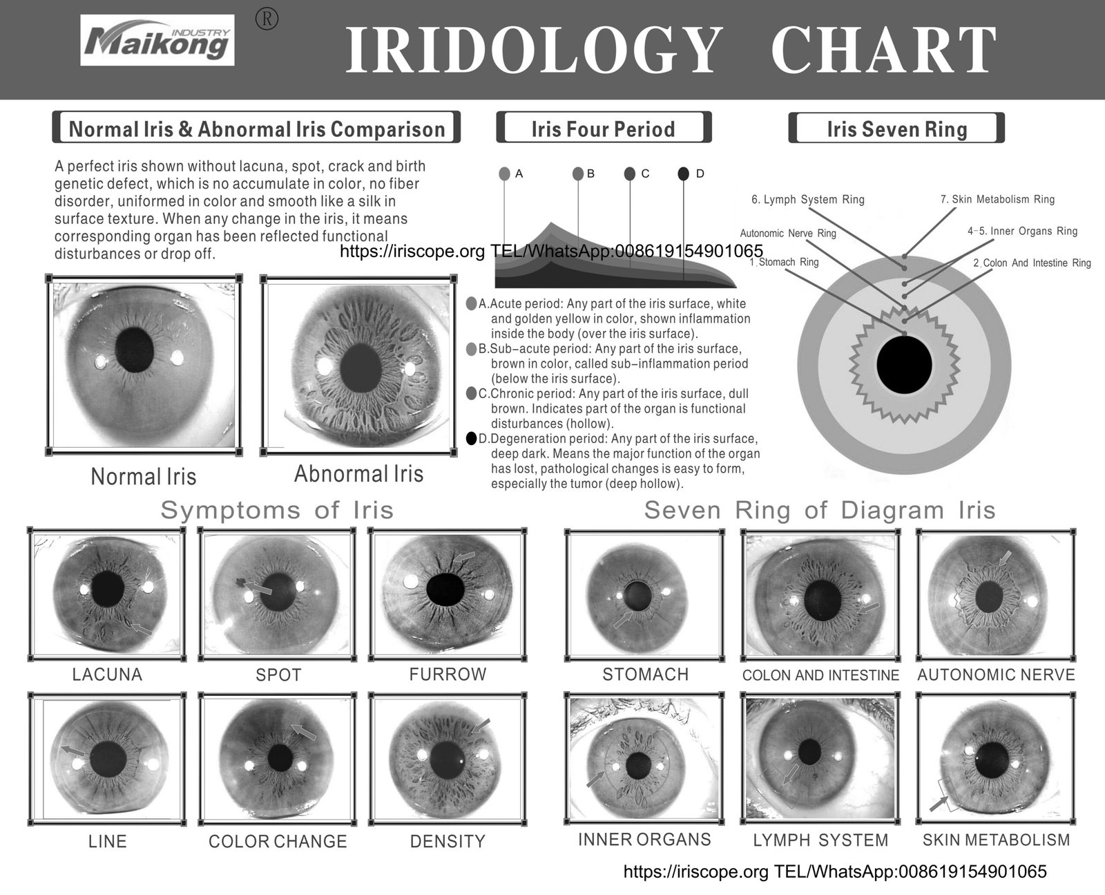
Step 3: Recognize Common Iris Signs
Learn to identify the most significant markings and what they may indicate:
• Nerve Rings: Circular rings that may indicate stress or nervous tension

• Lacunae: Enclosed darkened areas that may suggest inherent weaknesses

• Radii Solaris: Spoke-like lines radiating from the pupil that may indicate toxicity

• Pigmentation: Colored spots that may represent chemical deposits or inflammation

• Lymphatic Rosary: White cloudlike formations in the outer iris that may indicate lymphatic congestion

• Scurf Rim: Dark ring around the outer edge that may suggest skin elimination issues
Step 4: Assess Iris Color and Constitution
Examine the base iris color to determine the constitutional type, which may indicate general tendencies:
• Blue Iris (Lymphatic): May indicate sensitivity to respiratory and lymphatic issues

• Brown Iris (Hematogenic): May suggest stronger digestive function but potential blood sugar sensitivity

• Mixed Iris (Biliary): Combination of blue and brown, may indicate liver and digestive sensitivities

• Color variations and patterns provide additional information about specific organ conditions
Step 5: Document and Track Changes
For ongoing assessment, document observations and monitor for changes over time:
• Take clear photographs of both irises

• Note specific markings and their locations using clock positions

• Record color variations and intensity

• Compare with follow-up examinations to track changes

• Correlate observations with other health assessments and symptoms
Master Iridology Analysis
Get our comprehensive guide with detailed explanations of all iris signs and their potential health correlations.
Download Complete Guide
Scientific Perspective on Iridology Charts

Medical researcher reviewing scientific literature on iridology’s validity
The scientific community maintains a skeptical stance toward iridology as a diagnostic method. Understanding both the criticisms and the responses from iridology advocates provides important context for anyone exploring this practice.
Research Findings and Clinical Studies
Several controlled studies have examined iridology’s diagnostic accuracy with mixed results. A frequently cited 1979 study published in the Journal of the American Medical Association found that iridologists could not reliably detect gallbladder disease by examining photographs of irises. Similar studies testing iridology’s ability to identify kidney disease, cancer, and other conditions have generally not supported its diagnostic claims when subjected to blinded experimental conditions.
Proponents argue that these studies often fail to account for the holistic nature of iridology, which they maintain is not designed to diagnose specific diseases but rather to identify constitutional weaknesses and tissue changes. They also suggest that iridology works best as part of a comprehensive assessment rather than as a standalone diagnostic tool.
Physiological Basis and Theoretical Concerns

Anatomical illustration of eye structure and nervous system pathways
From a physiological perspective, conventional medicine acknowledges that certain systemic conditions can manifest visible changes in the eye. For example, diabetes may cause retinal changes, and liver disease can cause scleral yellowing. However, the specific iris-to-organ mappings proposed in iridology charts lack established neurological or vascular pathways that would explain how distant organ conditions could consistently alter specific iris segments.
Critics point out that iris pigmentation and structure are largely determined by genetics and remain relatively stable throughout life, with changes primarily related to local eye conditions rather than distant organ pathologies. Iridology practitioners counter that subtle changes in iris fibers, colors, and patterns do occur and can be documented with high-resolution imaging.
Balanced Perspective for Consumers
For individuals interested in iridology, a balanced approach recognizes both its limitations and potential value:
- Iridology should not replace conventional medical diagnosis or delay appropriate treatment
- It may have value as a complementary assessment tool within a holistic health framework
- The practice can encourage preventative health measures and lifestyle improvements
- Individual experiences with iridology vary widely and should be evaluated critically
- Qualified practitioners should acknowledge limitations and work cooperatively with medical professionals
This scientific context helps consumers make informed decisions about incorporating iridology into their health practices, understanding both its theoretical foundations and the current limitations in its evidence base.
5 Best Practices for Iridology Chart Interpretation

Professional iridologist demonstrating proper examination techniques and chart reference
1. Use Proper Equipment and Lighting
Accurate iris analysis requires appropriate tools and conditions. Professional iridologists use specialized equipment including magnifying lenses with at least 10x magnification, proper full-spectrum lighting, and often digital cameras with macro capabilities. Attempting to perform iris analysis with inadequate equipment leads to misinterpretations and missed details. Natural daylight or full-spectrum lighting that mimics natural light provides the most accurate color rendering for proper assessment.
2. Consider the Whole Person
Effective iridology never isolates iris signs from the individual’s complete health picture. Responsible practitioners gather comprehensive health history, consider current symptoms, and recognize that iris markings represent tendencies rather than definitive diagnoses. This contextual approach prevents overinterpretation of iris signs and acknowledges that genetic factors, environmental influences, and lifestyle choices all contribute to health outcomes beyond what may be visible in the iris.
3. Recognize Constitutional Types
Iris constitution—the genetic blueprint reflected in base iris color and structure—provides the foundation for accurate chart interpretation. Blue irises (lymphatic type), brown irises (hematogenic type), and mixed irises (biliary type) each suggest different inherent strengths and tendencies. Constitutional assessment helps practitioners understand the individual’s baseline and interpret other iris signs within this context. This approach acknowledges genetic predispositions while avoiding deterministic conclusions about health outcomes.
4. Document Baseline and Changes
Professional iridology practice involves establishing a baseline record and monitoring for changes over time. High-resolution photographs allow for comparison during follow-up assessments, helping to distinguish between permanent constitutional markings and potentially changeable signs. This documentation process supports the identification of improving or deteriorating conditions and provides objective evidence of changes that correlate with lifestyle modifications or therapeutic interventions.
5. Maintain Ethical Boundaries
Ethical iridology practice requires clear boundaries regarding diagnostic claims and treatment recommendations. Responsible practitioners explicitly communicate that iridology is a complementary assessment tool, not a replacement for medical diagnosis. They avoid making definitive disease diagnoses, discourage abandoning conventional medical care, and maintain appropriate referral relationships with healthcare providers. This ethical approach prioritizes client welfare while acknowledging the complementary role of iridology within a broader health framework.

Ethical practitioner explaining iridology’s scope and limitations to a client
By adhering to these best practices, iridology practitioners can provide more reliable and ethical services while clients can better evaluate the quality of iridology consultations they receive. These guidelines help maintain iridology as a potentially valuable complementary assessment tool while minimizing misuse or overreliance on its findings.
Enhance Your Holistic Health Knowledge
Sign up for our free mini-course on integrative approaches to health assessment, including iridology fundamentals.
Enroll in Free Mini-Course
Conclusion: The Place of Iridology Charts in Health Assessment
Iridology charts represent a fascinating intersection of ancient observational medicine and modern holistic health practices. As we’ve explored, these detailed iris maps offer alternative health practitioners a framework for assessing potential constitutional strengths, weaknesses, and tendencies through careful examination of iris structures, colors, and markings.
While scientific validation remains limited and conventional medicine maintains skepticism toward iridology’s diagnostic claims, the practice continues to evolve with improved technology, standardized approaches, and more nuanced applications. When approached with appropriate boundaries and ethical considerations, iridology may serve as one component of a comprehensive health assessment that encourages preventative care and personalized wellness strategies.
For those interested in exploring iridology, education remains essential—understanding both its potential contributions and its limitations helps individuals make informed decisions about incorporating this practice into their health journey. Whether viewed as a valuable assessment tool or simply a fascinating window into traditional healing approaches, iridology charts continue to intrigue those seeking diverse perspectives on health and wellness.
Continue Your Iridology Education
Download our comprehensive guide to understanding iridology charts and their application in holistic health assessment.
Get Your Complete Guide


