Iridology defined
Iridology defined
Iridology is defined as the science and study of the color and structure of the iris to determine tissue weakness and the body’s predisposition to weakness. The science of Iridology is NOT used for diagnosis. It is a tool used as a means of assessment for conditions and levels of health. The beginnings of Iridology have been cited from many areas of the world and date back to the time of the early Chaldeans (1000 B.C.) The first documented reference to iris analysis can be credited to the physician Philippus Meyens, who wrote a book called “Chiromatica Medica”, published in 1670, which described the reflexive features of the iris. In 1786, Christaen Haertels published his “De Oculo et Signo” translated meaning ‘the eye and its signs” and with this the significance of eye signs was gaining credence. The eye has been examined for diagnostic purposes for centuries. There are a number of references to the significance of bloodshot eyes in the Hippocratic writings, for instance, and the general color and brightness of the eyes have always been taken into account in orthodox examinations of sick patients. The theory that the iris can give more precise information about disease was first independently propounded about a century ago by a Hungarian, Ignatz von Peczely, and a Swede, Nils Liljequist. In 1881 von Peczely published a book on the Iris of the eye called “Discovery in the Realm of Nature and the Art of Healing”. Liljequist, also a discoverer of the rolle of the iris and its marking, published a two-volume work which was translated into Engilsh and called “Diagnosis from the Eye”. Other Iridologists such as Dr. J. Kritzer is known for his work, “Iris Diagnosis and Guide in Treatment”, and Peter Theil, of Germany, was recognized as the greatest Iridologist of his day. Dr. Henry Lahn, one of Lilequist’s students, brought the practice of Iridology to America near the turn of the century. In America, a chiropractor, Dr. Bernard Jensen, is hailed as the most accomplished Iridologist of recent years and is known for his healing philosophy and writings in the area of Iridology. A German Iridologist, Joseph Deck, has witten two full-color volumes, “Principles of Iris Diagnosis”, and Differentiation of Iris Marking”, both translated into English, complete with some of the most significant photo analysis. Physicians in Russia, Germany and France are more acquainted with iridological technique than those who practice in North America. In terms of science, the experimentation in iris evaluation is relatively new and should not be used in isolation but as a comprehensive system coupled with a medical history and other findings.

Essential elements to look for in best iridology camera
Essential elements to look for in best iridology camera
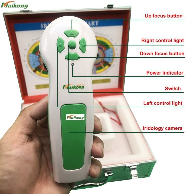
Our cold lighting fibre optic system minimizes client irritation and gives precise daylight illumination for optimum exposure and true colour every time. It includes variable intensity adjustment to enable even the darkest eyes to display every detail with no loss of contrast! In addition, our lights avoid the pupils to avoid putting artifacts in this vital assessment area.
Easy operatation. Get perfect focus with ease.
One of the most important features to consider is the lighting. Our fixed lighting system ensures easy comparative analysis. Side lighting option included.
Our designer has had more than 30 years clinical experience as an ND and Iridologist bringing with him a keen understanding of what professional Iridologists need.
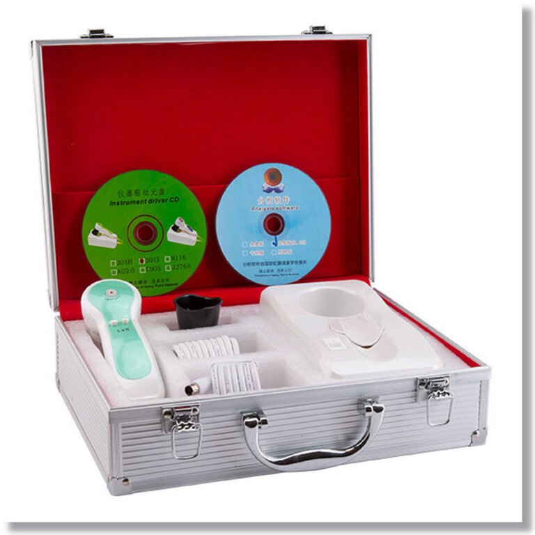
We only use high quality current model cameras and professional grade materials. Most of our camera systems include state of the art analysis software in the price.
All of our cameras meet iridology photographic criteria. In fact, we lead the way in iris photography!

Conclusion for iridology
Conclusion for iridology:
Iridology is an excellent example of pseudoscience in medicine, displaying many of the core features. It was invented by one individual based upon a single observation and emerging from a culture of quackery and pseudoscience. It follows a pre-scientific notion of biology – the homunculus model. It lacks any basis in anatomy, physiology, or any other basic science. Its practitioners are mostly “alternative” practitioners who use the technique as a cold reading. And the research clearly shows that iridology has absolutely no effect – it does not provide any useful information at all.
Anyone using or promoting iridology is, therefore, a pseudoscientific practitioner. Any profession that endorses iridology is not science-based and should be looked upon with suspicion.

What is iridology?
What is iridology?
Iridology (also known as iridodiagnosis or iridiagnosis) is an alternative medicine technique whose proponents claim that patterns, colors, and other characteristics of the iris can be examined to determine information about a patient’s systemic health. Practitioners match their observations to iris charts, which divide the iris into zones that correspond to specific parts of the human body. Iridologists see the eyes as “windows” into the body’s state of health.
Iridologists claim they can use the charts to distinguish between healthy systems and organs in the body and those that are overactive, inflamed, or distressed. Iridologists claim this information demonstrates a patient’s susceptibility towards certain illnesses, reflects past medical problems, or predicts later health problems.
As opposed to evidence-based medicine, iridology is not supported by quality research studies and is widely considered pseudoscience. The features of the iris are one of the most stable features on the human body throughout life.[disputed – discuss] The stability of iris structures is the foundation of the biometric technology which uses iris recognition for identification purposes.
In 1979, Bernard Jensen, a leading American iridologist, and two other iridology proponents failed to establish the basis of their practice when they examined photographs of the eyes of 143 patients in an attempt to determine which ones had kidney impairments. Of the patients, forty-eight had been diagnosed with kidney disease, and the rest had normal kidney function. Based on their analysis of the patients’ irises, the three iridologists could not detect which patients had kidney disease and which did not.
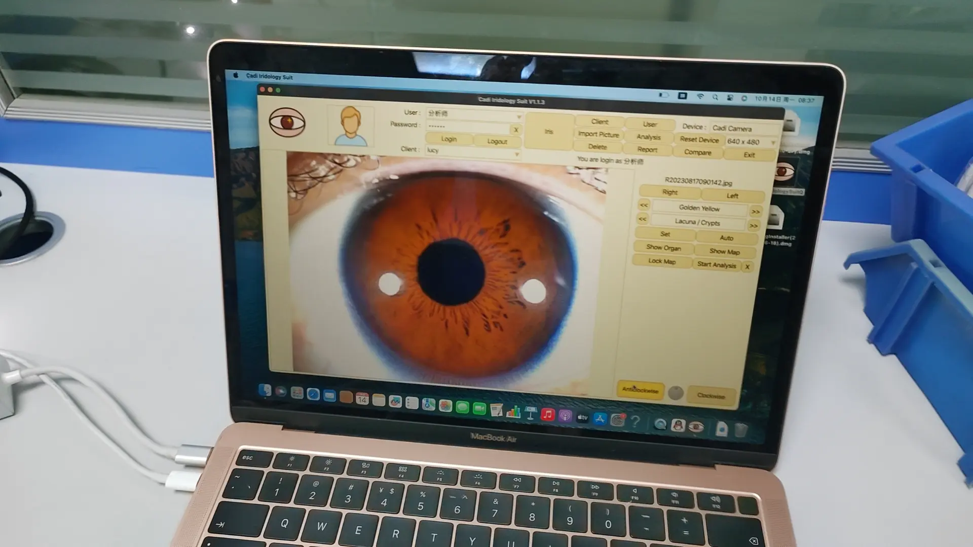
The iris is the greenish-yellow area surrounding the transparent pupil (showing as black). The white outer area is the sclera, the central transparent part of which is the cornea.Iridologists generally use equipment such as a flashlight and magnifying glass, cameras or slit-lamp microscopes to examine a patient’s irises for tissue changes, as well as features such as specific pigment patterns and irregular stromal architecture. The markings and patterns are compared to an iris chart that correlates zones of the iris with parts of the body. Typical charts divide the iris into approximately 80–90 zones. For example, the zone corresponding to the kidney is in the lower part of the iris, just before 6 o’clock. There are minor variations between charts’ associations between body parts and areas of the iris.
According to iridologists, details in the iris reflect changes in the tissues of the corresponding body organs. One prominent practitioner, Bernard Jensen, described it thus: “Nerve fibers in the iris respond to changes in body tissues by manifesting a reflex physiology that corresponds to specific tissue changes and locations.” This would mean that a bodily condition translates to a noticeable change in the appearance of the iris, but this has been disproven through many studies. (See section on Scientific research.) For example, acute inflammatory, chronic inflammatory and catarrhal signs may indicate involvement, maintenance, or healing of corresponding distant tissues, respectively. Other features that iridologists look for are contraction rings and Klumpenzellen, which may indicate various other health conditions.
Seven rings Iris zones/rings and accompanying body region/organ
Inner: Stomach.
Second: Small and large intestine.
Third: Circulation of blood and lymph.
Fourth: Internal organs and endocrine system.
Fifth: Musculoskeletal system.
Outer: Skin and organs of elimination.


