The human iris contains intricate patterns as unique as fingerprints. For practitioners of alternative medicine, these patterns represent more than just eye color—they’re a map to your health. An Iridologie-Diagramm serves as the key to decoding this map, linking specific zones of your iris to organs and systems throughout your body. Whether you’re curious about alternative health assessments or seeking to understand the colorful diagrams you’ve seen in natural health clinics, this comprehensive guide will explain how Iridologie-Diagramme work, their historical development, and the ongoing debate about their effectiveness.

Was ist ein Iridologie-Diagramm and How Does It Work?
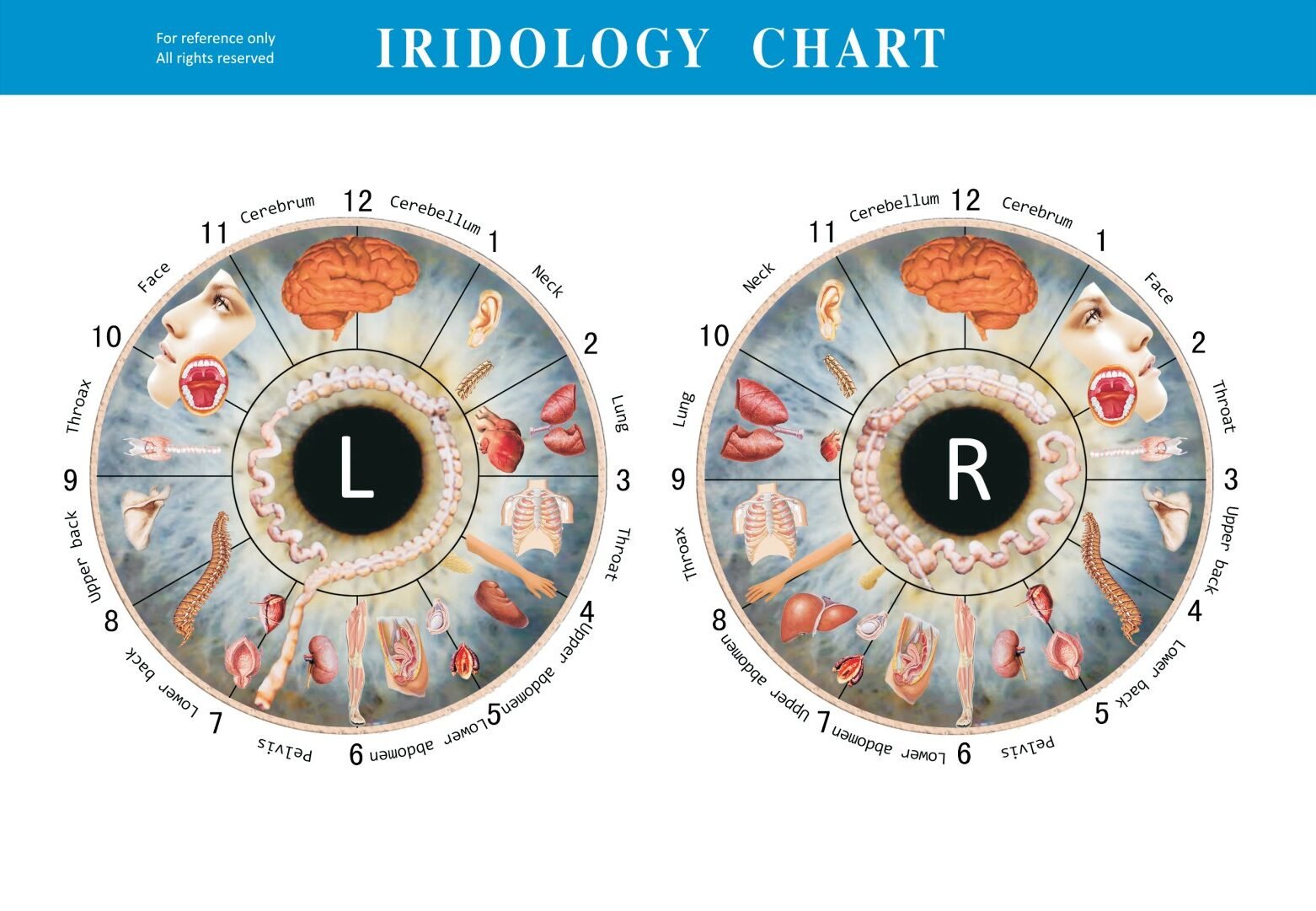
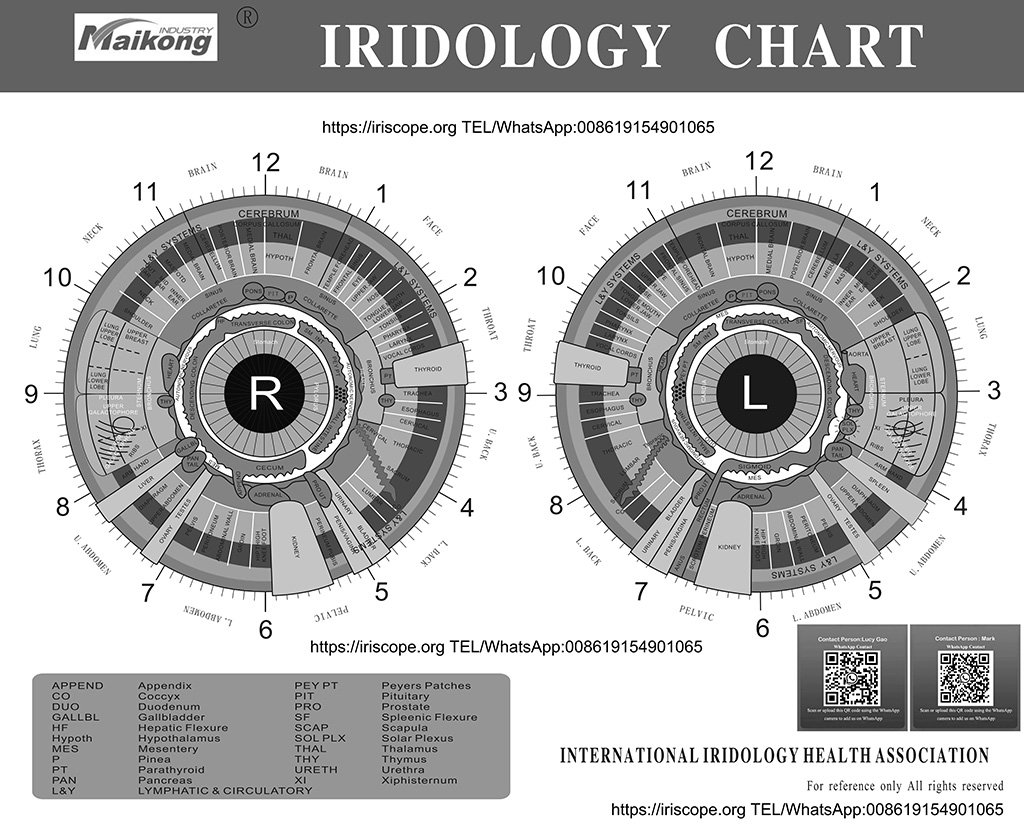
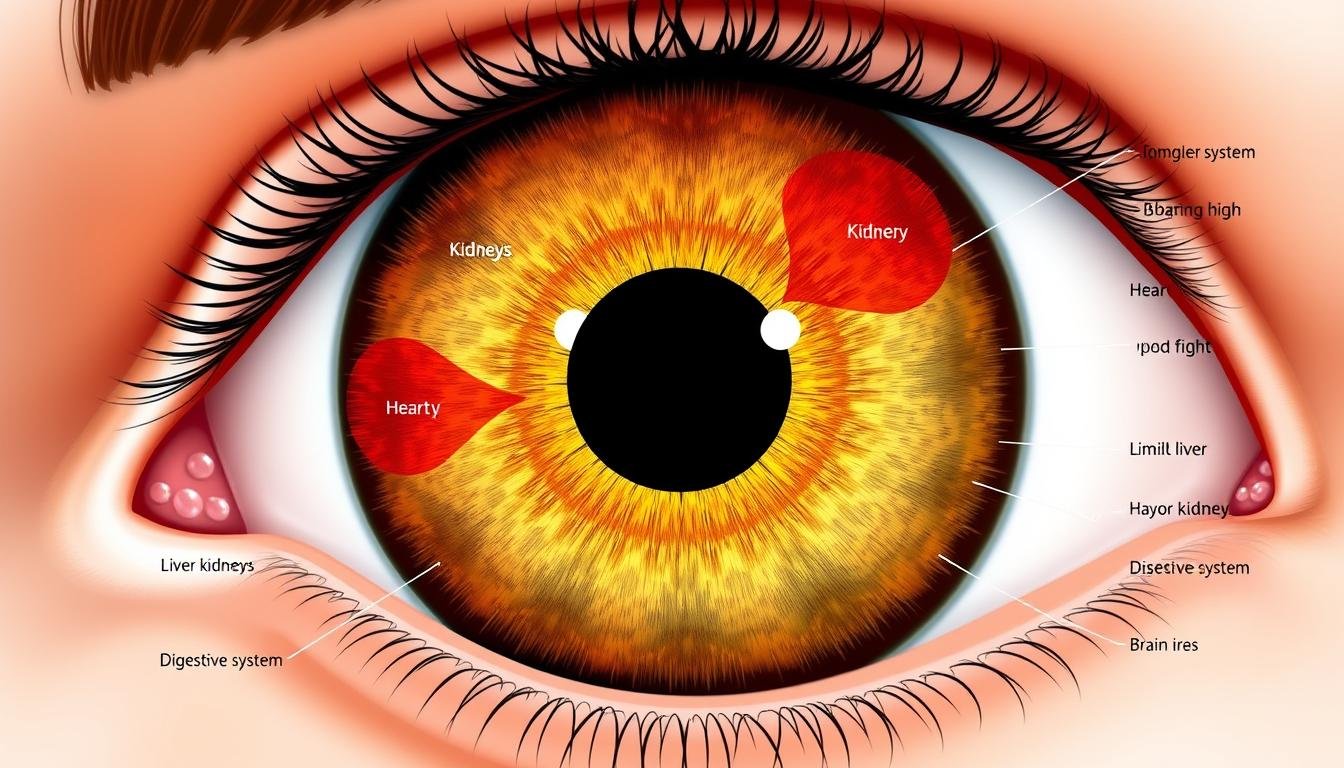
Ein Iridologie-Diagramm is a diagnostic tool that divides the iris into zones corresponding to different parts of the human body. Iridologists use these charts to examine the patterns, colors, and markings in your iris to assess your health status, identify potential problem areas, and even predict future health concerns.
Ein Standard Iridologie-Diagramm mapping iris zones to corresponding body organs and systems
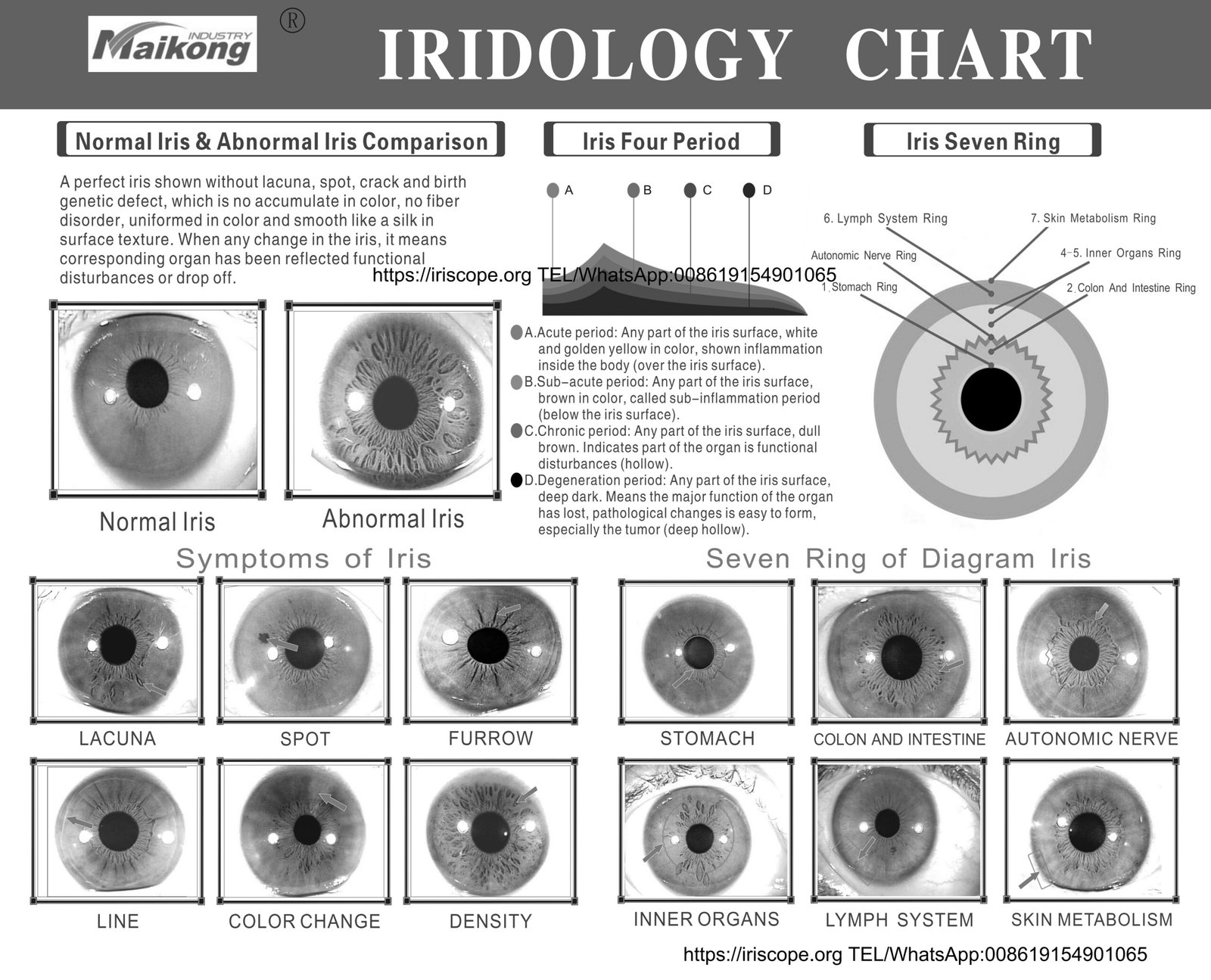
According to iridology practitioners, the iris connects to every organ and tissue via the nervous system and brain. When an organ experiences stress or damage, corresponding changes appear in the iris. These changes may manifest as:
- Color variations (white, yellow, brown spots)
- Structural changes (lines, rings, clouds)
- Texture alterations (density, fiber patterns)
- Pigmentation shifts (darkening or lightening)
By consulting an Iridologie-Diagramm, practitioners claim to identify which body systems need attention, even before symptoms appear elsewhere in the body.
The Historical Development of the Iridologie-Diagramm
The practice of examining the eyes for health insights dates back thousands of years, but modern Iridologie-Diagramme have a more recent history.


An early Iridologie-Diagramm from the 19th century
Key Figures in Iridologie-Diagramm Entwicklung
Philippus Meyeus (1670): Published the first explicit description of iridological principles in “Chiromatica Medica,” though he didn’t use the term “iridology.”
Ignaz von Peczely (1860s): The Hungarian physician is considered the founding father of modern iridology. Legend claims he noticed iris changes in an owl with a broken leg, inspiring his research. He created the first comprehensive Iridologie-Diagramm.
Nils Liljequist (1893): After observing changes in his own iris following medication, this Swedish homeopath published an atlas with 258 black and white illustrations and 12 color illustrations of the iris.
Bernard Jensen (1950s): Popularized iridology in the United States through classes and publications. His Iridologie-Diagramm remains influential in modern practice.
Over time, Iridologie-Diagramme have evolved from simple drawings to detailed, color-coded maps. Modern charts often incorporate digital technology for more precise analysis, though the fundamental principles remain similar to those established by the early pioneers.
Understanding Iris Zones on an Iridologie-Diagramm
Ein Standard Iridologie-Diagramm divides the iris into approximately 80-90 zones, arranged in concentric circles around the pupil. Each zone corresponds to different parts of the body.

Concentric zones on an Iridologie-Diagramm representing different body systems
Major Zones of the Iridologie-Diagramm
Richtige Iriszonen
- Top Region: Brain, head, sinuses
- Upper Right: Lungs, bronchi, throat
- Upper Left: Heart, chest, breast
- Middle Right: Leber, Gallenblase
- Middle Left: Stomach, pancreas
- Lower Region: Intestines, reproductive organs
Verließ Iriszonen
- Top Region: Brain, cerebral circulation
- Upper Right: Spleen, left shoulder
- Upper Left: Heart, thyroid
- Middle Right: Stomach, pancreas
- Middle Left: Adrenals, kidneys
- Lower Region: Colon, bladder, reproductive system
Circular Zones
- Pupillary Zone: Digestive system
- Ciliary Zone: Muscular system, circulation
- Autonomic Nerve Wreath: Nervensystem
- Outer Iris Zone: Skin, extremities, lymphatic system
- Iris Edge: Skeletal system
Iridologists examine these zones for color changes, structural marks, and other anomalies. The position of a mark within a zone further refines the diagnosis to specific organs or tissues.

Comparison of healthy iris patterns (left) versus patterns indicating potential health issues (right) according to iridology
Organs and Systems Mapped in the Iridologie-Diagramm
Ein Iridologie-Diagramm maps virtually every organ and system in the human body to specific locations on the iris. Here’s how some key organs are represented:

Major organ systems as mapped on a standard Iridologie-Diagramm
| Organ/System | Iris Location | Signs of Potential Issues |
| Herz | Left iris, upper quadrant (7-8 o’clock) | Dark spots, white cloudiness, radial lines |
| Liver | Right iris, middle area (8-9 o’clock) | Brown spots, yellowish discoloration |
| Kidneys | Lower quadrants of both irises (5-6 o’clock) | White marks, dark spots, structural changes |
| Lunge | Upper quadrants of both irises (2-3 o’clock) | White clouds, dark spots, structural breaks |
| Verdauungssystem | Around the pupil in both irises | Discolorations, radial lines, dark spots |
| Lymphsystem | Outer edge of both irises | White marks, cloudiness, structural changes |
Iridologists believe that examining these specific areas can reveal information about organ function, inflammation, toxicity, and overall health status. The intensity and type of markings may indicate the severity of conditions.
Interested in Learning More?
Discover how iridology practitioners interpret these organ mappings with our comprehensive guide.
Kostenlose Beratung anfordern
Health Conditions Identified Through an Iridologie-Diagramm
Proponents of iridology claim that various health conditions can be detected by analyzing iris patterns and comparing them to an Iridologie-Diagramm. While scientific validation remains limited, here are some conditions that iridologists commonly look for:

Various iris markings and their potential health indications according to iridology
Circulatory Conditions
- Hypertension: Indicated by a ring around the iris, suggesting increased blood pressure.
- High Cholesterol: A white ring around the outer edge of the iris may indicate excessive cholesterol or arteriosclerosis.
- Poor Circulation: Shown by a lack of clear definition in iris fibers.
Verdauungsprobleme
- Liver Damage: Brown spots in the liver area may indicate liver disease or toxicity.
- Gallbladder Problems: Yellowing in specific areas may suggest gallbladder or bile duct abnormalities.
- Digestive Weakness: Discolorations around the pupil can indicate problems with the digestive system.
Other Conditions
- Inflammation: White streaks or clouds in specific zones may indicate inflammation in corresponding organs.
- Weakened Immune System: White marks throughout the iris may suggest compromised immunity.
- Hormonal Imbalances: Specific markings in endocrine system zones may indicate thyroid or adrenal issues.
“While iridology cannot diagnose specific diseases, it may help identify which organs and systems in the body are under stress and at risk of developing conditions.”
Iridologists emphasize that their practice is not meant to diagnose diseases but rather to identify areas of weakness or stress in the body. They often recommend further medical testing to confirm any suspected conditions.
Scientific Perspective on the Iridologie-Diagramm
The scientific community has expressed significant skepticism about the validity of Iridologie-Diagramme and the practice of iridology in general. Understanding both sides of this debate is important for anyone considering iridology.

Wissenschaftliche Forschung zur Prüfung der Gültigkeit iridologischer Behauptungen
Argumente zur Unterstützung der Iridologie
- Die Iris enthält Tausende von Nervenenden, die mit dem Gehirn verbunden sind
- Anecdotal evidence from practitioners reporting successful assessments
- Long historical use in various traditional medicine systems
- Non-invasive nature makes it an appealing complementary approach
- Kann vorbeugende Gesundheitsmaßnahmen und Selbstbewusstsein fördern
Wissenschaftliche Kritik
- Controlled studies have failed to demonstrate diagnostic accuracy
- Iris structure is genetically determined and remains stable throughout life
- No proven physiological mechanism for how internal organs would affect iris appearance
- Practitioners often show poor inter-examiner reliability
- May delay proper medical diagnosis and treatment
Schlüsselforschungsergebnisse
Several controlled studies have tested iridology’s effectiveness:
- 1979 Study: Bernard Jensen and two other iridologists failed to correctly identify patients with kidney disease by examining iris photographs.
- 1980s British Medical Journal Study: Iridologists could not correctly identify patients with gallbladder disease from iris examinations.
- 2000 Review by Edzard Ernst: Found no benefit from iridology in controlled, masked evaluations of diagnostic validity.
- 2005 Cancer Detection Study: Concluded iridology was of no value in diagnosing common forms of cancer.
The Australian Government’s Department of Health reviewed alternative therapies in 2015 and found no clear evidence of effectiveness for iridology.
Despite these criticisms, many practitioners and patients continue to find value in iridology as a complementary approach to health assessment, particularly when used alongside conventional medical care rather than as a replacement.
How to Read a Basic Iridologie-Diagramm
If you’re interested in exploring iridology, understanding how to read an Iridologie-Diagramm is the first step. While professional training is recommended for accurate interpretation, here are the basics:

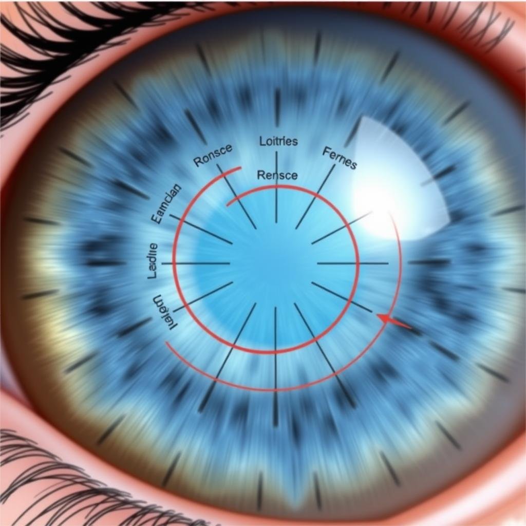
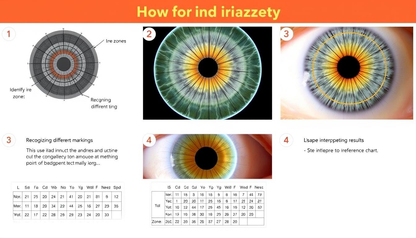
Step-by-step guide to basic Iridologie-Diagramm reading
Step-by-Step Guide to Reading an Iridologie-Diagramm
Schritt 1: Identifizieren Sie den IRIS -Typ
Determine whether you’re looking at a blue, brown, mixed, or other iris type. Different base colors may require slightly different interpretations.
Step 2: Orient the Chart
Position the Iridologie-Diagramm correctly—right iris corresponds to the right side of the body, left iris to the left side.
Step 3: Locate the Zones
Identify the major zones (pupillary, ciliary, etc.) and the organ-specific regions within each iris.
Step 4: Analyze Markings
Look for color changes, structural marks, and other anomalies in each zone and compare to reference materials.
Häufige Iriszeichen und ihre Bedeutung
| Iris Zeichen | Aussehen | Möglicher Hinweis |
| Radiale Linien | Lines extending from pupil toward iris edge | Nerve stress or irritation in corresponding organ |
| Lacuna | Geschlossene, seeartige Formationen | Inherent weakness or genetic predisposition |
| Kryptionen | Offene, grubenartige Formationen | Potential for toxin accumulation |
| Natriumring | Weißer Ring um die äußere Iris | Possible salt/mineral imbalance |
| Lymphatischer Rosenkranz | Weiße Punkte bilden einen Kreis | Verstopfung des Lymphsystems |
| Schorffelge | Dark ring at outer edge of iris | Probleme mit der Hautentfernung |
Wichtig: Self-diagnosis using an Iridologie-Diagramm is not recommended. Always consult with qualified health professionals for any health concerns.

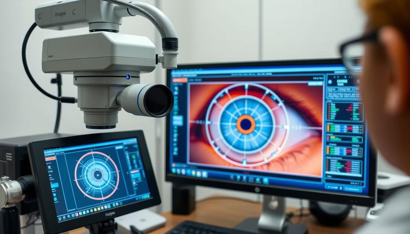
A professional iridologist examining a patient’s iris using specialized equipment
Conclusion: The Value and Limitations of the Iridologie-Diagramm
Der Iridologie-Diagramm represents a fascinating intersection of traditional observation and alternative health assessment. While scientific evidence does not currently support many of iridology’s claims, the practice continues to attract interest from those seeking holistic approaches to health.

Modern digital approaches to iridology analysis
For those interested in iridology, it’s important to approach it with a balanced perspective:
- Consider it a complementary tool rather than a replacement for conventional medical diagnosis
- Consult with qualified practitioners who have proper training
- Maintain healthy skepticism while remaining open to potential insights
- Always follow up on significant health concerns with appropriate medical testing
Whether you view the Iridologie-Diagramm as a valuable health assessment tool or an interesting historical artifact of alternative medicine, understanding its principles and limitations allows you to make informed decisions about its role in your health journey.


